Copyright
©The Author(s) 2022.
World J Clin Cases. Jun 16, 2022; 10(17): 5620-5633
Published online Jun 16, 2022. doi: 10.12998/wjcc.v10.i17.5620
Published online Jun 16, 2022. doi: 10.12998/wjcc.v10.i17.5620
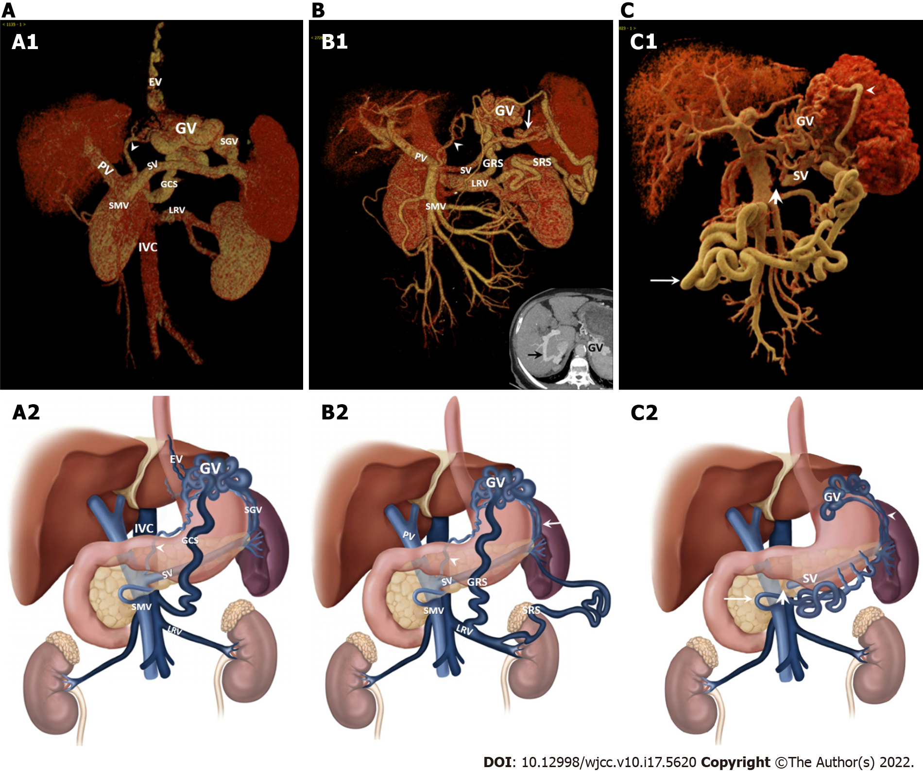
Figure 2 Computed tomography portal venography of gastric variceal collateral vessels.
A: Coronal oblique volume-rendered (VR) computed tomography (CT) portal venogram views (A1) and schematic drawing (A2) illustrated collateral circulation of esophageal varices (GVs) in the patient with gastroesophageal varices (75-years-old male patients with liver cirrhosis). GVs were supplied by left gastric vein (LGV) (arrowhead) and SGV, and drained by gastrocaval shunt (GCS), and esophageal and para-esophageal varices (EVs); B: Coronal oblique VR CT portal venogram views (B1) and schematic drawing (B2) illustrated collateral circulation of GVs in the patient with isolated gastric varices (IGV1) (75-years-old female patients). GVs were supplied by LGV (arrowhead) and SGV (white arrow), and drained by GRS, SRS and intrahepatic portosystemic shunts (black arrow in the MIP image); C: Coronal oblique cinematically rendered reconstruction in CT portal venogram views (C1) and schematic drawing (C2) showing collateral vessels in a 42-years-old male patient with IGV1 caused by pancreatic pseudocyst secondary to pancreatitis. GVs were supplied by SGV (arrowhead), spleno-gastroomental -superior mesenteric shunt (white arrow) was a major collateral vessel due to partial splenic vein occlusion (thick arrow).
- Citation: Song YH, Xiang HY, Si KK, Wang ZH, Zhang Y, Liu C, Xu KS, Li X. Difference between type 2 gastroesophageal varices and isolated fundic varices in clinical profiles and portosystemic collaterals. World J Clin Cases 2022; 10(17): 5620-5633
- URL: https://www.wjgnet.com/2307-8960/full/v10/i17/5620.htm
- DOI: https://dx.doi.org/10.12998/wjcc.v10.i17.5620


