Copyright
©The Author(s) 2022.
World J Clin Cases. Jun 6, 2022; 10(16): 5352-5358
Published online Jun 6, 2022. doi: 10.12998/wjcc.v10.i16.5352
Published online Jun 6, 2022. doi: 10.12998/wjcc.v10.i16.5352
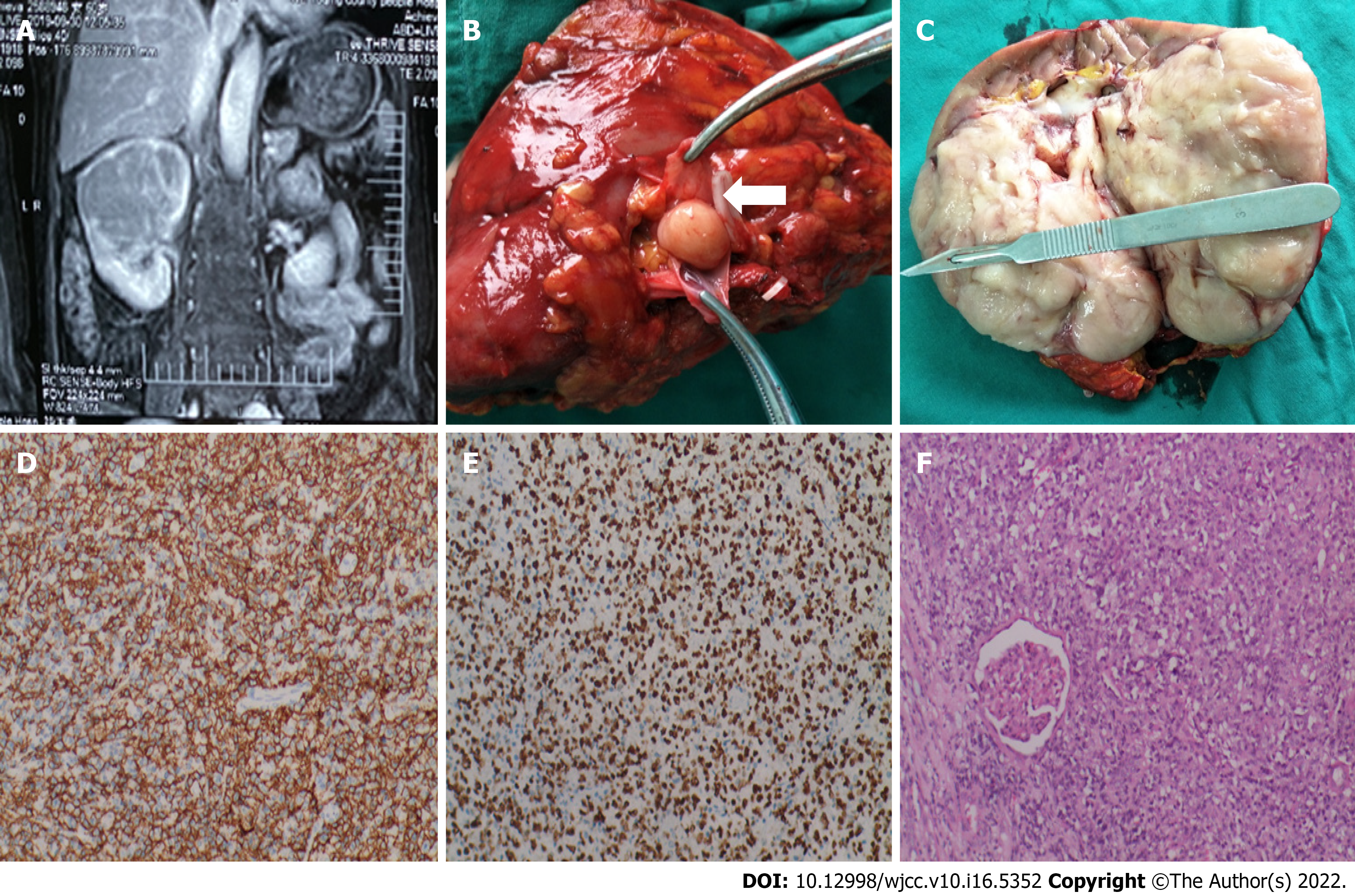
Figure 1 Imaging and pathological data.
A: Magnetic resonance imaging revealed a space-occupying lesion of the right kidney; B: The tumor thrombus extended along the right renal vein (arrow); C: The gross specimen was a total kidney with partial ureteral resection; D and E: Immunohistochemical staining positive was for CD20 and positive for Ki-67; F: The tumor cells showed medium-to-large lymphoid cells infiltrating the growth.
- Citation: He J, Mu Y, Che BW, Liu M, Zhang WJ, Xu SH, Tang KF. Comprehensive treatment for primary right renal diffuse large B-cell lymphoma with a renal vein tumor thrombus: A case report. World J Clin Cases 2022; 10(16): 5352-5358
- URL: https://www.wjgnet.com/2307-8960/full/v10/i16/5352.htm
- DOI: https://dx.doi.org/10.12998/wjcc.v10.i16.5352


