Copyright
©The Author(s) 2022.
World J Clin Cases. May 26, 2022; 10(15): 5103-5110
Published online May 26, 2022. doi: 10.12998/wjcc.v10.i15.5103
Published online May 26, 2022. doi: 10.12998/wjcc.v10.i15.5103
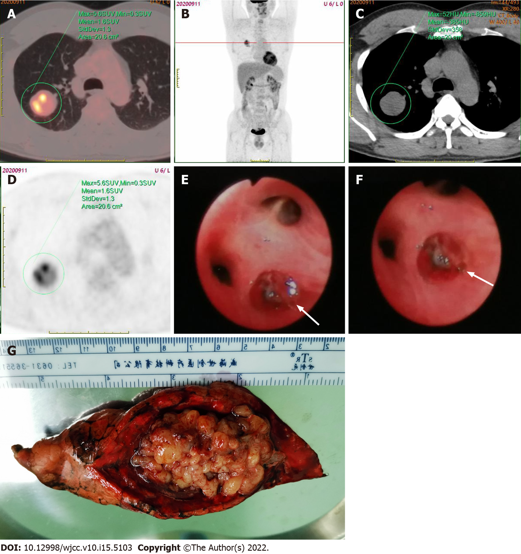
Figure 2 Positron emission tomography-computed tomography scan.
A-D: Showed a solitary round mass in the upper lobe of the right lung, with unevenly increased radioactivity uptake and an SUVmax of 5.6; E and F: Fiberoptic bronchoscopy revealed the presence of new organisms (white arrows) at the posterior opening of the upper right lobe, with a smooth capsule, surrounded by purulent secretions, and oozing a small amount of blood; G: The specimen obtained following the thoracoscopic resection of the posterior segment of the right upper lobe of the lung was grayish yellow in color, with some small round-like changes and a soft texture.
- Citation: He WW, Huang ZX, Wang WJ, Li YL, Xia QY, Qiu YB, Shi Y, Sun HM. Solitary primary pulmonary synovial sarcoma: A case report. World J Clin Cases 2022; 10(15): 5103-5110
- URL: https://www.wjgnet.com/2307-8960/full/v10/i15/5103.htm
- DOI: https://dx.doi.org/10.12998/wjcc.v10.i15.5103


