Copyright
©The Author(s) 2022.
World J Clin Cases. May 26, 2022; 10(15): 5051-5056
Published online May 26, 2022. doi: 10.12998/wjcc.v10.i15.5051
Published online May 26, 2022. doi: 10.12998/wjcc.v10.i15.5051
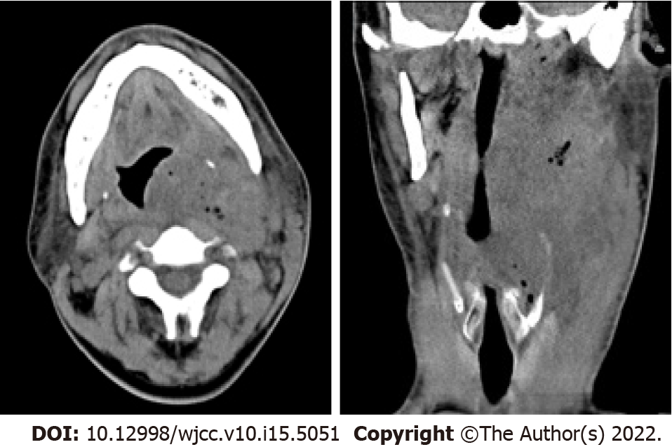
Figure 1 Plain computed tomography revealed a soft tissue mass containing scattered air (dimensions: 9.
9 cm × 7.3 cm × 4.6 cm; computed tomography value: 22-34 HU) that had a vague boundary and an irregular shape on the left neck and submaxillary space, and the oropharynx and laryngeal left lateral wall were thickened.
- Citation: Xie TH, Zhao WJ, Li XL, Hou Y, Wang X, Zhang J, An XH, Liu LT. Carotid blowout syndrome caused by chronic infection: A case report. World J Clin Cases 2022; 10(15): 5051-5056
- URL: https://www.wjgnet.com/2307-8960/full/v10/i15/5051.htm
- DOI: https://dx.doi.org/10.12998/wjcc.v10.i15.5051


