Copyright
©The Author(s) 2022.
World J Clin Cases. May 26, 2022; 10(15): 5012-5017
Published online May 26, 2022. doi: 10.12998/wjcc.v10.i15.5012
Published online May 26, 2022. doi: 10.12998/wjcc.v10.i15.5012
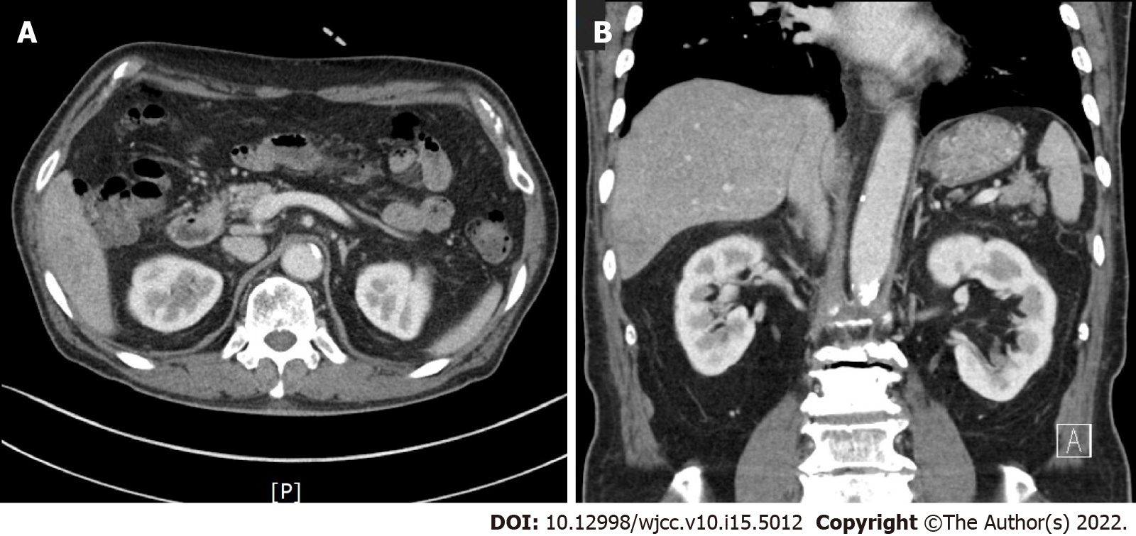
Figure 4 Follow-up abdominal computed tomography after antibiotic treatment for 33 d showing resolution of the pyogenic liver abscess.
A and B: Axial and coronal images showing resolution of the focal ill-defined low attenuation in liver segment 6.
- Citation: Park JG, Suh JI, Kim YU. Gastric heterotopia of colon found cancer workup in liver abscess: A case report. World J Clin Cases 2022; 10(15): 5012-5017
- URL: https://www.wjgnet.com/2307-8960/full/v10/i15/5012.htm
- DOI: https://dx.doi.org/10.12998/wjcc.v10.i15.5012


