Copyright
©The Author(s) 2022.
World J Clin Cases. May 26, 2022; 10(15): 4991-4997
Published online May 26, 2022. doi: 10.12998/wjcc.v10.i15.4991
Published online May 26, 2022. doi: 10.12998/wjcc.v10.i15.4991
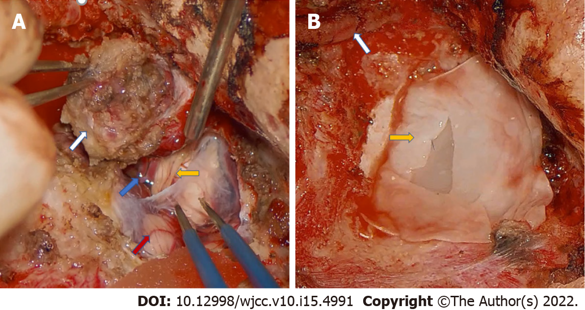
Figure 4 Intraoperative pictures of the second stage surgery.
A: The tumor (white arrow) that invaded the meninges; The anterior inferior cerebellar artery (blue arrow); The cochlear nerve (yellow arrow); The cerebellum (red arrow); B: The artificial dura mater (yellow arrow) used to repair the dura of the posterior fossa; The internal carotid artery (white arrow).
- Citation: Zhao Y, Zhao Y, Zhang LQ, Feng GD. Postoperative infection of the skull base surgical site due to suppurative parotitis: A case report. World J Clin Cases 2022; 10(15): 4991-4997
- URL: https://www.wjgnet.com/2307-8960/full/v10/i15/4991.htm
- DOI: https://dx.doi.org/10.12998/wjcc.v10.i15.4991


