Copyright
©The Author(s) 2022.
World J Clin Cases. May 26, 2022; 10(15): 4971-4984
Published online May 26, 2022. doi: 10.12998/wjcc.v10.i15.4971
Published online May 26, 2022. doi: 10.12998/wjcc.v10.i15.4971
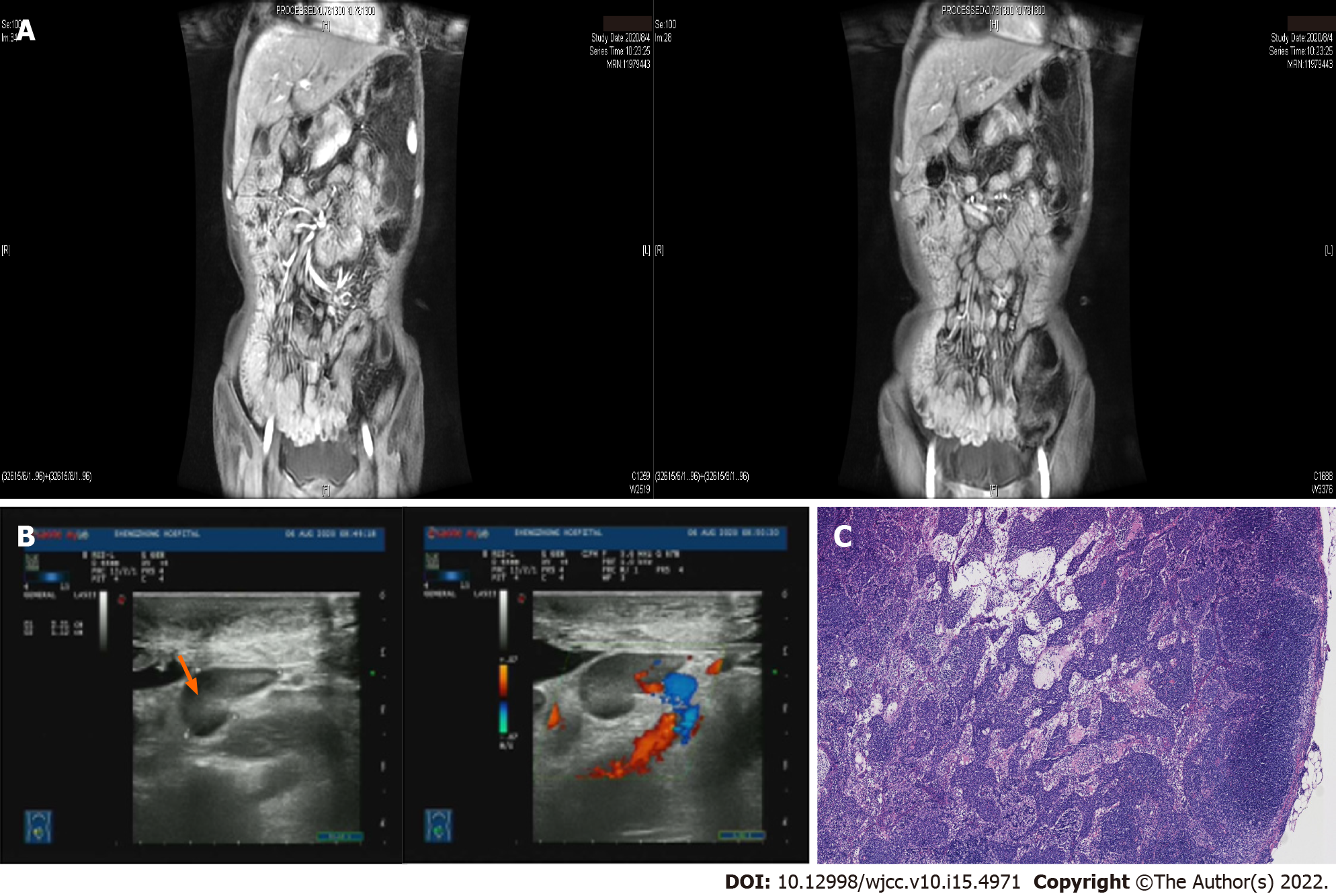
Figure 1 Magnetic resonance imaging and retroperitoneal B-ultrasound manifestations.
A: Thickened small bowel wall and whole colon, with enlarged regional lymph nodes at the mesenteries; B: Retroperitoneal B-ultrasound manifestation: Multiple retroperitoneal lymph nodes are enlarged. The orange arrow points to the largest swollen lymph node; C: Pathological results of lymph node puncture showing the destruction of lymph node structure and diffuse proliferation and infiltration of tumor cells in the paracortical area and medullary sinus.
- Citation: Weng CY, Ye C, Fan YH, Lv B, Zhang CL, Li M. CD8-positive indolent T-Cell lymphoproliferative disorder of the gastrointestinal tract: A case report and review of literature. World J Clin Cases 2022; 10(15): 4971-4984
- URL: https://www.wjgnet.com/2307-8960/full/v10/i15/4971.htm
- DOI: https://dx.doi.org/10.12998/wjcc.v10.i15.4971


