Copyright
©The Author(s) 2022.
World J Clin Cases. May 16, 2022; 10(14): 4704-4708
Published online May 16, 2022. doi: 10.12998/wjcc.v10.i14.4704
Published online May 16, 2022. doi: 10.12998/wjcc.v10.i14.4704
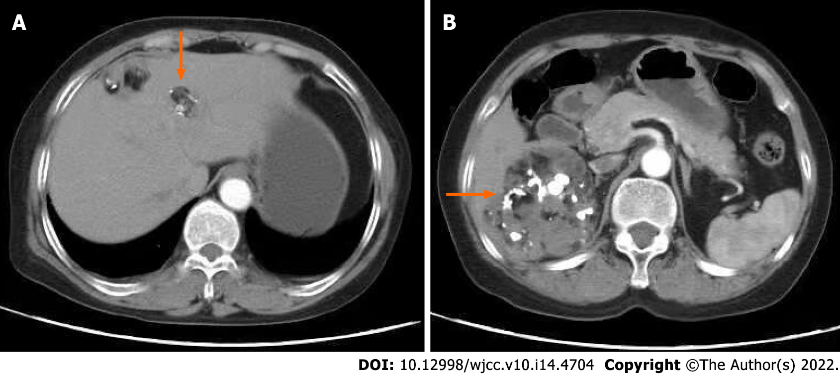
Figure 1 Abdominal computed tomography showed multiple masses in the abdominal cavity.
A: Showing three small masses in the liver; B: Showed the largest one was located in the posterior peritoneum next to the sixth segment of the right liver. The largest one was located in the posterior peritoneum next to the sixth segment of the right liver, three masses were present inside of liver and one mass was in the right pelvic floor.
- Citation: Hu X, Jia Z, Zhou LX, Kakongoma N. Ovarian growing teratoma syndrome with multiple metastases in the abdominal cavity and liver: A case report. World J Clin Cases 2022; 10(14): 4704-4708
- URL: https://www.wjgnet.com/2307-8960/full/v10/i14/4704.htm
- DOI: https://dx.doi.org/10.12998/wjcc.v10.i14.4704


