Copyright
©The Author(s) 2022.
World J Clin Cases. May 16, 2022; 10(14): 4632-4639
Published online May 16, 2022. doi: 10.12998/wjcc.v10.i14.4632
Published online May 16, 2022. doi: 10.12998/wjcc.v10.i14.4632
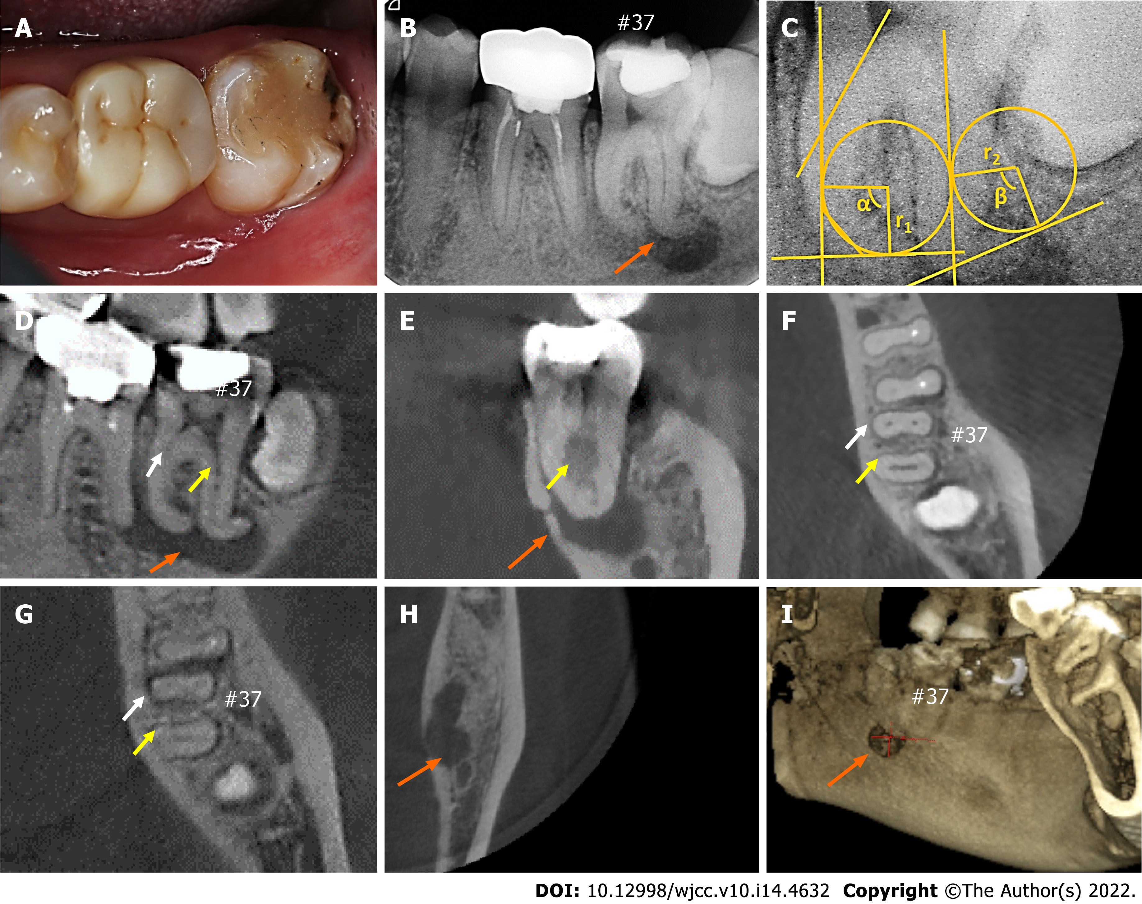
Figure 1 Initial clinical situation (#37).
A: Photograph of the mandibular second molar; B: Preoperative periapical radiograph of the molar; C: Measurement of the radius of the curvature and angles; D and E: Sagittal (D) and coronal (E) dimensions obtained from cone beam computed tomography (CBCT); F-H: Axial dimensions obtained from CBCT; I: Three-dimensional reconstruction of CBCT images presenting the perforation of the lingual cortical plate. White and yellow arrows represent the mesial root canals and a distal root canal, respectively; orange arrows show the regions of large periapical radiolucency.
- Citation: Xu LJ, Zhang JY, Huang ZH, Wang XZ. Successful individualized endodontic treatment of severely curved root canals in a mandibular second molar: A case report. World J Clin Cases 2022; 10(14): 4632-4639
- URL: https://www.wjgnet.com/2307-8960/full/v10/i14/4632.htm
- DOI: https://dx.doi.org/10.12998/wjcc.v10.i14.4632


