Copyright
©The Author(s) 2022.
World J Clin Cases. May 16, 2022; 10(14): 4541-4549
Published online May 16, 2022. doi: 10.12998/wjcc.v10.i14.4541
Published online May 16, 2022. doi: 10.12998/wjcc.v10.i14.4541
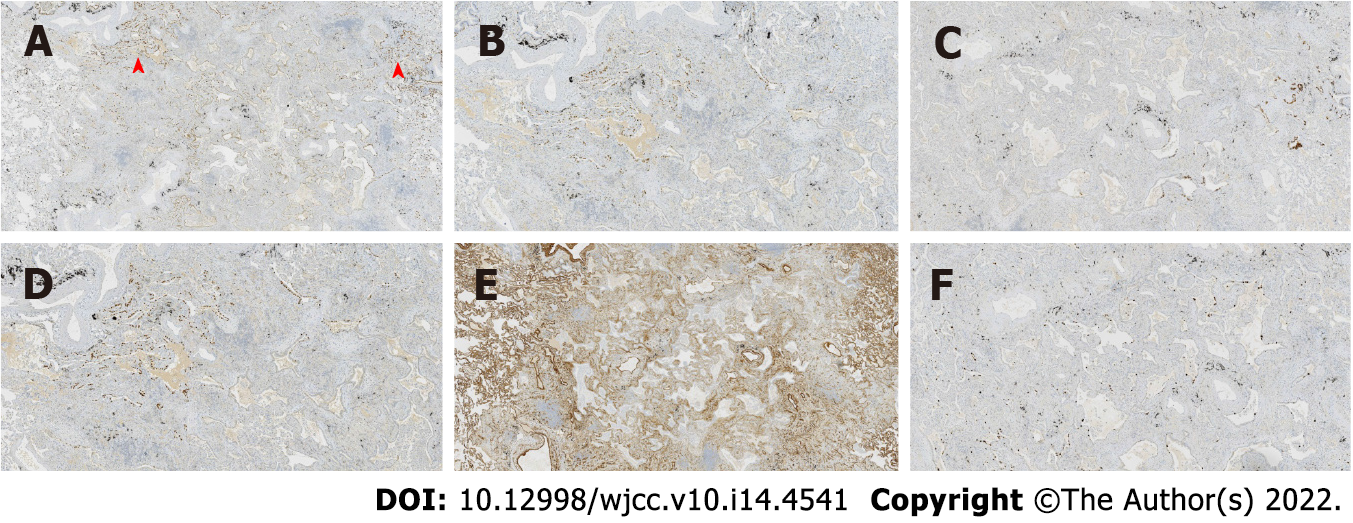
Figure 3 Immunohistochemical staining in case 1.
Thyroid transcription factor 1 was expressed in bronchioles and the surrounding tumor glands, with the only difference being intensity. The results of P40, P63, and cytokeratin 5/6 staining were the same, and positive staining was detected only in the bilayer structures of the tumor. Collagen IV staining showed the presence of alveolar structure, and the Ki-67 index was low. A: Thyroid transcription factor 1; B: P40; C: Cytokeratin 5/6; D: P63; E: Collagen IV; F: Ki-67.
- Citation: Du Y, Wang ZY, Zheng Z, Li YX, Wang XY, Du R. Bronchiolar adenoma with unusual presentation: Two case reports. World J Clin Cases 2022; 10(14): 4541-4549
- URL: https://www.wjgnet.com/2307-8960/full/v10/i14/4541.htm
- DOI: https://dx.doi.org/10.12998/wjcc.v10.i14.4541


