Copyright
©The Author(s) 2022.
World J Clin Cases. May 16, 2022; 10(14): 4541-4549
Published online May 16, 2022. doi: 10.12998/wjcc.v10.i14.4541
Published online May 16, 2022. doi: 10.12998/wjcc.v10.i14.4541
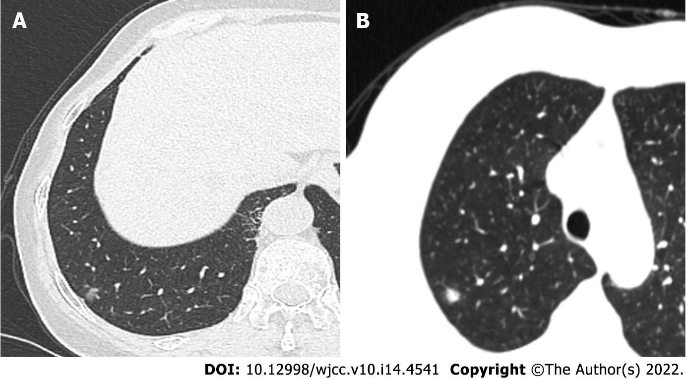
Figure 1 Imaging findings of pulmonary nodules in case 1 and case 2.
A: A mixed ground-glass nodule with a diameter of 0.6 cm in the subpleura of the posterior basal segment of the lower lobe of the right lung in case 1. The texture was relatively uniform, and the nodule was slightly pulled near the pleura; B: A solid lobulated nodule at the apex of the right upper lobe, 8 mm in diameter, with blurred edge, in case 2.
- Citation: Du Y, Wang ZY, Zheng Z, Li YX, Wang XY, Du R. Bronchiolar adenoma with unusual presentation: Two case reports. World J Clin Cases 2022; 10(14): 4541-4549
- URL: https://www.wjgnet.com/2307-8960/full/v10/i14/4541.htm
- DOI: https://dx.doi.org/10.12998/wjcc.v10.i14.4541


