Copyright
©The Author(s) 2022.
World J Clin Cases. May 16, 2022; 10(14): 4509-4518
Published online May 16, 2022. doi: 10.12998/wjcc.v10.i14.4509
Published online May 16, 2022. doi: 10.12998/wjcc.v10.i14.4509
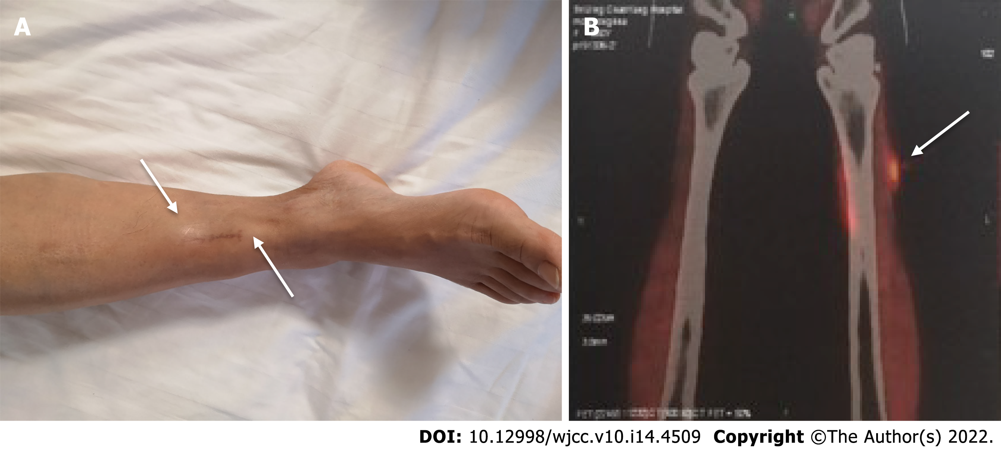
Figure 1 Appearance of nodules.
A: Clinical photograph showing two subcutaneous nodules on the right lower leg (white arrow); B: Positron emission tomography computed tomography scan showing increased fluorodeoxyglucose uptake in an anterolateral tibialis anterior nodule (white arrow).
- Citation: Liu Y, Zhu J, Huang YH, Zhang QR, Zhao LL, Yu RH. Cutaneous mucosa-associated lymphoid tissue lymphoma complicating Sjögren's syndrome: A case report and review of literature. World J Clin Cases 2022; 10(14): 4509-4518
- URL: https://www.wjgnet.com/2307-8960/full/v10/i14/4509.htm
- DOI: https://dx.doi.org/10.12998/wjcc.v10.i14.4509


