Copyright
©The Author(s) 2022.
World J Clin Cases. May 16, 2022; 10(14): 4470-4479
Published online May 16, 2022. doi: 10.12998/wjcc.v10.i14.4470
Published online May 16, 2022. doi: 10.12998/wjcc.v10.i14.4470
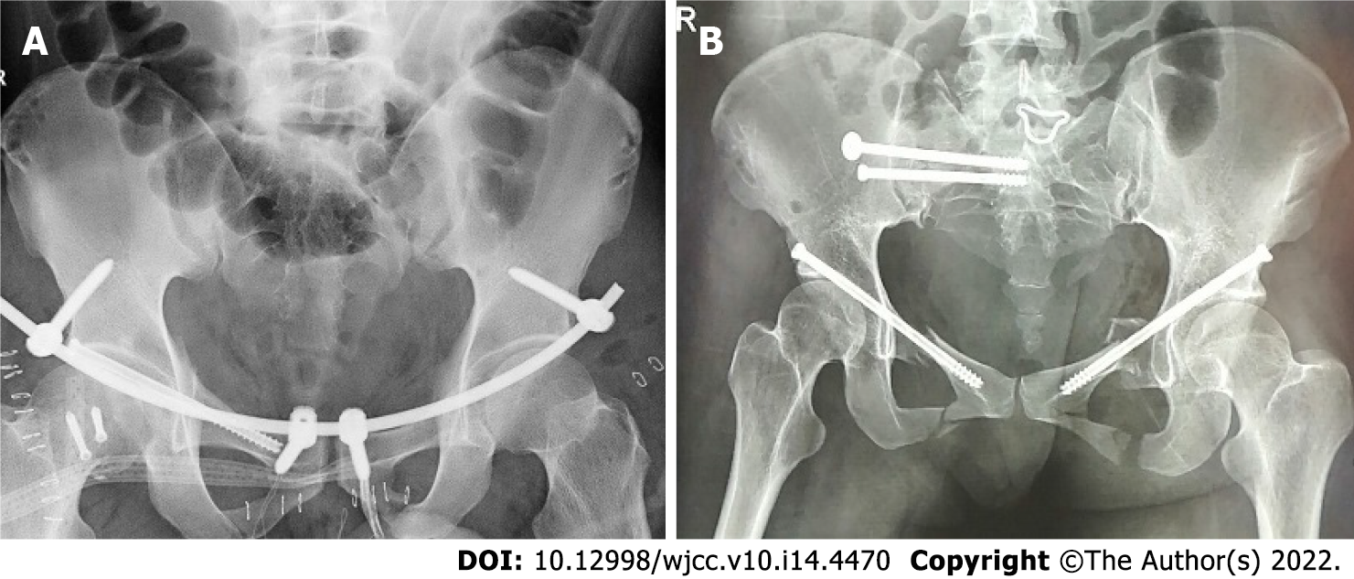
Figure 5 Postoperative pelvic X-ray images.
A, B: Pelvic X-ray (A) and multi-slice spiral computed tomography scan (B) were used to observe the pelvic reduction at 3 mo postoperative.
- Citation: Huang JG, Zhang ZY, Li L, Liu GB, Li X. Multi-slice spiral computed tomography in diagnosing unstable pelvic fractures in elderly and effect of less invasive stabilization. World J Clin Cases 2022; 10(14): 4470-4479
- URL: https://www.wjgnet.com/2307-8960/full/v10/i14/4470.htm
- DOI: https://dx.doi.org/10.12998/wjcc.v10.i14.4470


