Copyright
©The Author(s) 2022.
World J Clin Cases. May 6, 2022; 10(13): 4249-4263
Published online May 6, 2022. doi: 10.12998/wjcc.v10.i13.4249
Published online May 6, 2022. doi: 10.12998/wjcc.v10.i13.4249
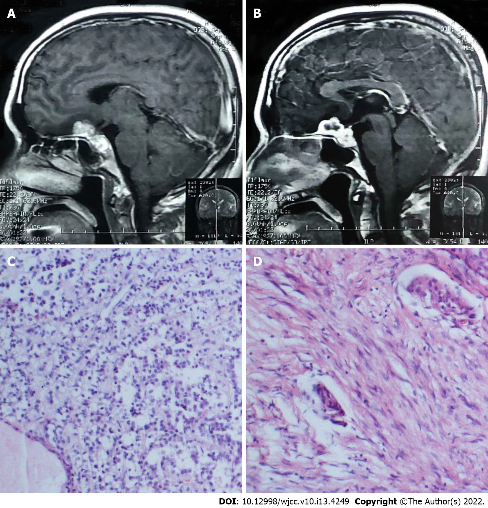
Figure 3 Histopathological examination showed a meningioma and non-functioning pituitary adenoma.
A and B: Mid-sagittal contrast magnetic resonance imaging showed space-occupying lesions located in the planum sphenoidale and sellar region; C: Histological examination revealed pituitary adenoma; D: Histological examination revealed meningioma.
- Citation: Hu TH, Wang R, Wang HY, Song YF, Yu JH, Wang ZX, Duan YZ, Liu T, Han S. Coexistence of meningioma and other intracranial benign tumors in non-neurofibromatosis type 2 patients: A case report and review of literature. World J Clin Cases 2022; 10(13): 4249-4263
- URL: https://www.wjgnet.com/2307-8960/full/v10/i13/4249.htm
- DOI: https://dx.doi.org/10.12998/wjcc.v10.i13.4249


