Copyright
©The Author(s) 2022.
World J Clin Cases. May 6, 2022; 10(13): 4249-4263
Published online May 6, 2022. doi: 10.12998/wjcc.v10.i13.4249
Published online May 6, 2022. doi: 10.12998/wjcc.v10.i13.4249
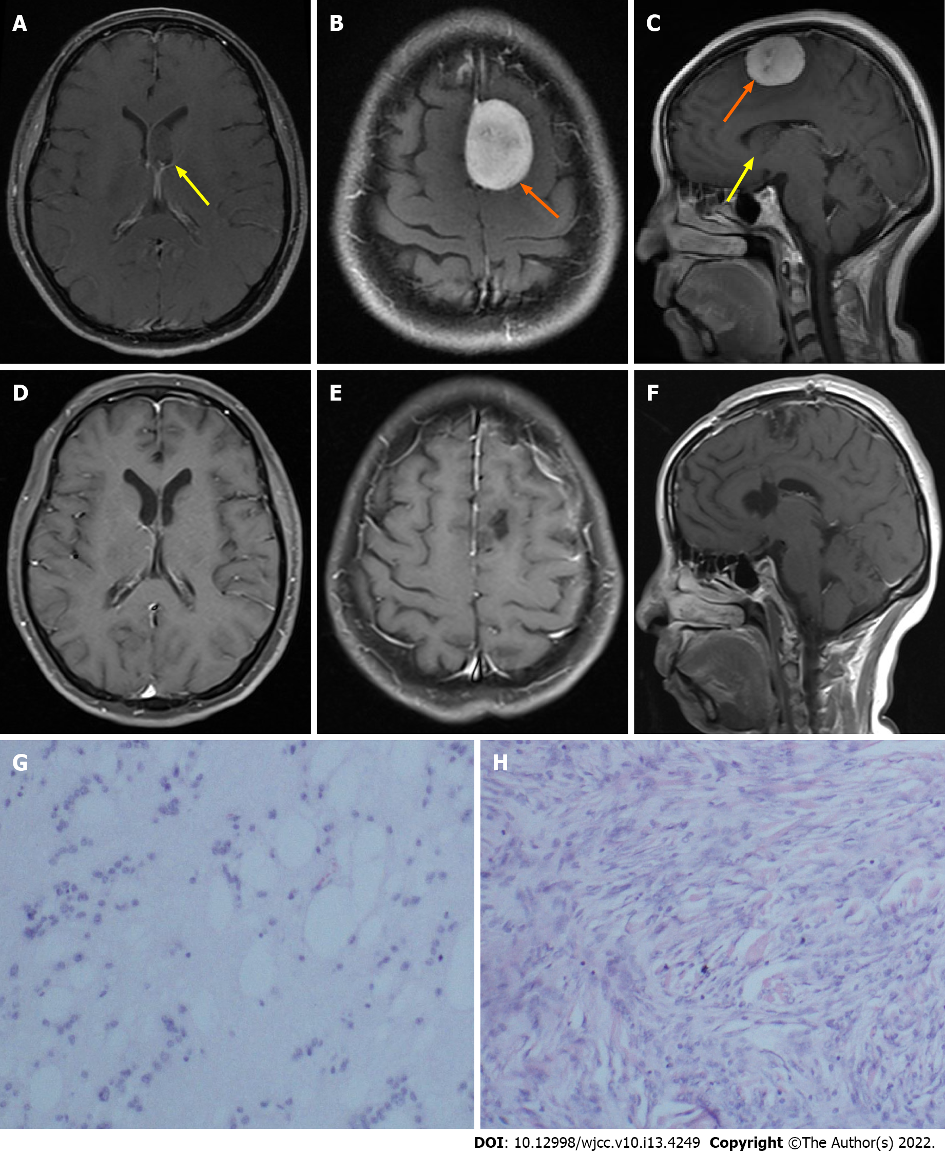
Figure 1 Histopathological examination showed a meningioma and subependymoma.
A-C: Preoperative axial and coronal contrast-enhanced magnetic resonance imaging (MRI) showed a mass in the left lateral ventricle (yellow arrow) and a mass in the left parietal parafalcine (orange arrow); D-F: Postoperative axial and coronal MRI showed that gross total resection of the tumors was achieved; G: Histological examination revealed subependymoma; H: Histological examination revealed meningioma.
- Citation: Hu TH, Wang R, Wang HY, Song YF, Yu JH, Wang ZX, Duan YZ, Liu T, Han S. Coexistence of meningioma and other intracranial benign tumors in non-neurofibromatosis type 2 patients: A case report and review of literature. World J Clin Cases 2022; 10(13): 4249-4263
- URL: https://www.wjgnet.com/2307-8960/full/v10/i13/4249.htm
- DOI: https://dx.doi.org/10.12998/wjcc.v10.i13.4249


