Published online May 6, 2022. doi: 10.12998/wjcc.v10.i13.4185
Peer-review started: September 13, 2021
First decision: October 18, 2021
Revised: October 27, 2021
Accepted: March 27, 2022
Article in press: March 27, 2022
Published online: May 6, 2022
Acute pancreatitis is an uncommon complication of gastrointestinal endoscopy, especially if the patient has none of the common risk factors associated with pancreatitis; such as alcoholism, gallstones, hypertriglyceridemia, hypercalcemia or the use of certain drugs.
A 56-year-old female patient developed abdominal pain immediately after the completion of an upper gastrointestinal endoscopy. The pain was predominantly in the upper and middle abdomen and was persistent and severe. The patient was diagnosed with acute pancreatitis. Treatment included complete fasting, octreotide injection prepared in a prefilled syringe to inhibit pancreatic enzymes secretion, ulinastatin injection to inhibit pancreatic enzymes activity, esome
Pancreatitis should be considered in the differential diagnosis of abdominal pain after upper and lower gastrointestinal endoscopy.
Core Tip: Acute pancreatitis is an uncommon complication of gastrointestinal endoscopy, especially when the patient has none of the common risk factors associated with pancreatitis; such as alcoholism, gallstones, hypertriglyceridemia, hypercalcemia or the use of certain drugs. We report an unusual case of acute pancreatitis related to gastrointestinal endoscopy. It is important to recognize this complication in order that appropriate treatment can be undertaken quickly for an optimal outcome.
- Citation: Dai MG, Li LF, Cheng HY, Wang JB, Ye B, He FY. Acute pancreatitis as a rare complication of gastrointestinal endoscopy: A case report. World J Clin Cases 2022; 10(13): 4185-4189
- URL: https://www.wjgnet.com/2307-8960/full/v10/i13/4185.htm
- DOI: https://dx.doi.org/10.12998/wjcc.v10.i13.4185
Endoscopy is a widely used diagnostic and therapeutic procedure and is usually well tolerated by patients. Potential complications include perforation, bleeding, postoperative polyps and side effects associated with sedation and analgesia[1-3]. Rare complications have also been reported in the literature including spleen trauma, infection, diverticulitis and appendicitis[4]. Acute pancreatitis is a well-documented complication of endoscopic retrograde cholangiopancreatography[5], but not as a complication of upper digestive endoscopy[6]. To our knowledge, only a few cases of acute pancreatitis as a complication of digestive endoscopy have been reported in the English literature. These cases were due to colonoscopy. Here, we report a case of acute pancreatitis as a rare complication after gastro
A 56-year-old woman underwent non-sedation gastrointestinal endoscopy for early cancer screening. It was the first gastrointestinal endoscopy for the patient. She had a sharp abdominal pain approximately 2 h after completion of the procedure once she had arrived home.
She presented with severe nausea and vomiting 2 h after the procedure. The patient did not have obvious abdominal pain immediately after the procedure. The pain was predominantly in the upper and middle abdomen, was persistent, severe and with no radiation. Pain was accompanied by nausea and non-projectile vomiting of stomach contents. Flatulence was reduced. The patient had a mild fever without chills, diarrhea, chest tightness, chest pain or any other discomfort.
Her past medical history included hepatitis B. She had no history of alcoholism, gallstones or pancreatitis.
Her birth history and feeding history were uneventful. There was no history of similar illness in the family.
On initial evaluation, vital signs revealed a temperature of 37.3°C, pulse rate of 77 bpm, blood pressure of 147/77 mmHg; and respiration rate of 15breaths/min. The patient was conscious and oriented. No yellowing of the skin or eyes was observed. Both lungs were clear, no dry or moist crackles (rales) were heard. The patient had tenderness in the upper and middle abdomen, no rebound pain or muscle tension was noted. Murphy’s sign, McBurney’s sign, and shifting dullness were all negative, and bowel sounds were heard at a rate of 3/min. No edema in the lower extremities was observed. No pathological signs were found.
Laboratory examination results were as follows: CRP 61.6 mg/L; white blood cells 15.5 x 10⁹ cells/L; amylase level 1022 IU/L (normal 23-184 IU/L); lipase level 4264 U/dL (normal 1-35 U/dL); arterial blood gas findings pH 7.36, HCO3 22 mmol/L; hepatobiliary enzyme and blood lipids were normal; serum calcium 2.0 mmol/L; hepatitis B (HB) surface antigen positive, HBeAg positive, HB core antibody positive; erythrocyte sedimentation rate 95 mm/h.
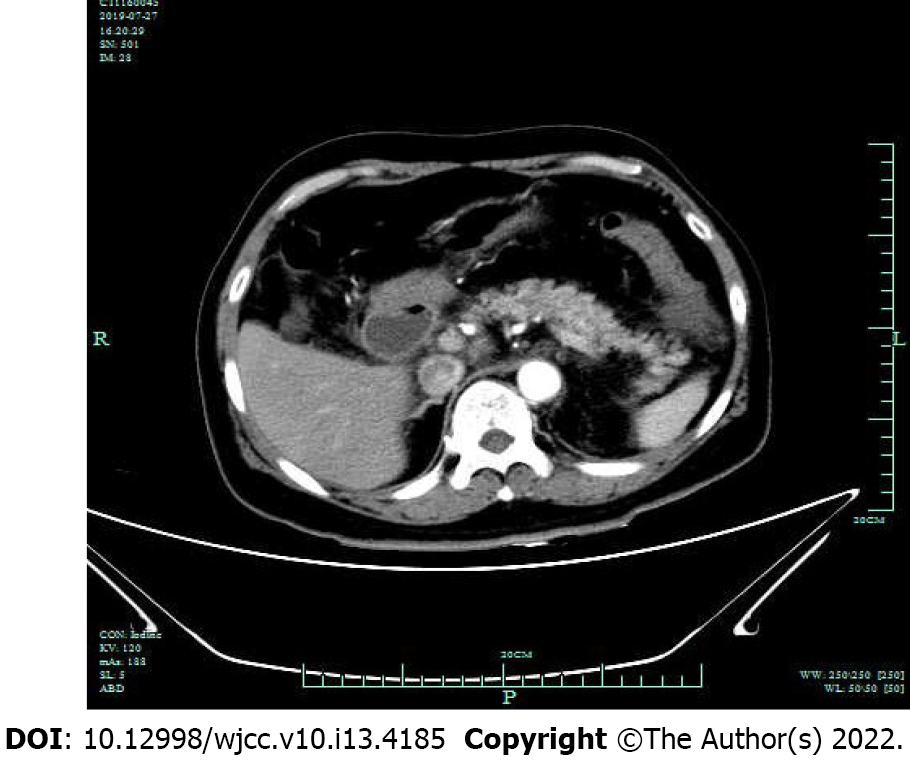
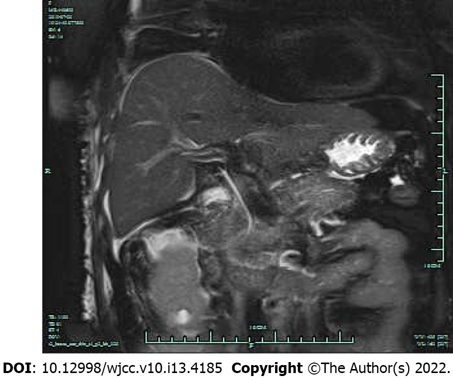
The patient's upper gastrointestinal endoscopy was normal. A contrast-enhanced abdominal computed tomography scan after admission suggested acute pancreatitis with peripancreatic fluid collection (Figure 1). Two incidental renal cysts and uterine fibroids were also detected. Magnetic resonance cholangiopancreatography revealed no structural changes and no gallstones in the pancreaticobiliary duct system (Figure 2).
Acute pancreatitis.
Treatment included complete fasting, octreotide injection prepared in a prefilled syringe to inhibit pancreatic enzymes secretion, ulinastatin injection to inhibit pancreatic enzymes activity, esomeprazole for gastric acid suppression, fluid replacement and nutritional support.
Over the next 3 d, the patient's symptoms improved, and serum amylase levels decreased to 104 IU/L within the normal range. The patient remained hemodynamically stable throughout hospitalization and was discharged home in a clinically stable state.
Although upper gastrointestinal endoscopy has not yet been demonstrated to be associated with an increased risk of pancreatitis and the relationship between endoscopy and pancreatitis may have been coincidental, both occurred within a short time and may explain the causality. In addition, the patient had no risk factors related to pancreatitis, such as alcoholism, trauma (including iatrogenic trauma), drugs, or infections[7]. Moreover, the patient had previously been tested for autoimmune pancreatitis, but the results were negative and lipid levels were normal. Therefore, we consider that gastrointestinal endoscopy may have played a role in the development of acute pancreatitis. In the literature, only one case of pancreatitis secondary to upper gastrointestinal endoscopy was reported in 1982[8]. This is the first case of pancreatitis secondary to gastrointestinal endoscopy reported in China.
Endoscopy is an essential procedure for gastroenterologists. The number and technical difficulties of endoscopies have increased over the past few decades and quality and safety remain important. The complication of pancreatitis caused by upper and lower gastrointestinal endoscopy is uncommon. Four cases of acute pancreatitis following upper and lower gastrointestinal endoscopy were considered to be caused by mechanical trauma due to manipulation of the colonoscope[6,9-11]. The potential mechanisms involved in the pathogenesis of pancreatitis include the following three factors: bile reflux due to high pressure[12]; mechanical trauma during the procedure[4,11,13]; and asymptomatic hyperamylasemia[14-17].
Since the development of acute necrotizing pancreatitis caused by upper gastrointestinal endoscopy has no relationship with previous pancreatic injury, the most probable etiology in this patient was severe vomiting and excessive pressure in the abdominal cavity, causing bile reflux into the pancreatic ducts, consequently activating trypsinogen to trypsin, which led to self-digestion of the pancreas. Bile reflux due to high pressure is considered an important cause of pancreatitis in clinical practice. In a previous study, hyperamylasemia was reported in 12% of patients undergoing endoscopy, but it was thought to be secondary to increased secretion of the salivary amylase isoenzyme[18]. Apart from the causes described above, we have been unable to find any other associations.
Whether it was a result of direct local trauma or an undetermined release of inflammatory mediators, clinically symptomatic acute pancreatitis is unusual among the complications of conventional endoscopic procedures. The diagnosis of acute pancreatitis is complex. It may be suspected clinically, but biochemical, radiological, and sometimes histological evidence is needed to confirm the diagnosis. Pancreatitis should be considered in the differential diagnosis of abdominal pain after upper and lower gastrointestinal endoscopy, when the most common explanations for such pain are excluded. Therefore, it is important to recognize this emergency in order that appropriate treatment can be undertaken for an optimal outcome.
Provenance and peer review: Unsolicited article; Externally peer reviewed.
Peer-review model: Single blind
Specialty type: Gastroenterology and hepatology
Country/Territory of origin: China
Peer-review report’s scientific quality classification
Grade A (Excellent): 0
Grade B (Very good): 0
Grade C (Good): C
Grade D (Fair): 0
Grade E (Poor): E
P-Reviewer: Ardengh JC, Brazil; Hirai R, Japan S-Editor: Ma YJ L-Editor: Filipodia P-Editor: Ma YJ
| 1. | Ghazi A, Grossman M. Complications of colonoscopy and polypectomy. Surg Clin North Am. 1982;62:889-896. [PubMed] [DOI] [Cited in This Article: ] |
| 2. | Vernava AM, Longo WE. Complications of endoscopic polypectomy. Surg Oncol Clin N Am. 1996;5:663-673. [PubMed] [Cited in This Article: ] |
| 3. | Palmer KR. Complications of gastrointestinal endoscopy. Gut. 2007;56:456-457. [PubMed] [DOI] [Cited in This Article: ] |
| 4. | Thomas AW, Mitre RJ. Acute pancreatitis as a complication of colonoscopy. J Clin Gastroenterol. 1994;19:177-178. [PubMed] [DOI] [Cited in This Article: ] |
| 5. | Bilbao MK, Dotter CT, Lee TG, Katon RM. Complications of endoscopic retrograde cholangiopancreatography (ERCP). A study of 10,000 cases. Gastroenterology. 1976;70:314-320. [PubMed] [Cited in This Article: ] |
| 6. | Nevins AB, Keeffe EB. Acute pancreatitis after gastrointestinal endoscopy. J Clin Gastroenterol. 2002;34:94-95. [PubMed] [DOI] [Cited in This Article: ] |
| 7. | Imrie CW, McKay AJ, Benjamin IS, Blumgart LH. Secondary acute pancreatitis: aetiology, prevention, diagnosis and management. Br J Surg. 1978;65:399-402. [PubMed] [DOI] [Cited in This Article: ] |
| 8. | Deschamps JP, Allemand H, Janin Magnificat R, Camelot G, Gillet M, Carayon P. Acute pancreatitis following gastrointestinal endoscopy without ampullary cannulation. Endoscopy. 1982;14:105-106. [PubMed] [DOI] [Cited in This Article: ] |
| 9. | Kulling D, Sahai AV, Knapple WL, Cunningham JT, Hoffman BJ. Diagnostic endoscopic ultrasound of the pancreas may cause acute pancreatitis. Endoscopy. 1998;30:S7-S8. [PubMed] [DOI] [Cited in This Article: ] |
| 10. | Limb C, Ibrahim IA, Fitzsimmons S, Harper AJ. Recurrent pancreatitis after unremarkable colonoscopy, temporalised by CT imaging: an unusual case. BMJ Case Rep. 2016;2016. [PubMed] [DOI] [Cited in This Article: ] |
| 11. | Ko HH, Jamieson T, Bressler B. Acute pancreatitis and ileus post colonoscopy. Can J Gastroenterol. 2009;23:551-553. [PubMed] [DOI] [Cited in This Article: ] |
| 12. | Fischer M, Hassan A, Sipe BW, Fogel EL, McHenry L, Sherman S, Watkins JL, Schmidt S, Lazzell-Pannell L, Lehman GA. Endoscopic retrograde cholangiopancreatography and manometry findings in 1,241 idiopathic pancreatitis patients. Pancreatology. 2010;10:444-452. [PubMed] [DOI] [Cited in This Article: ] |
| 13. | Khashram M, Frizelle FA. Colonoscopy--a rare cause of pancreatitis. N Z Med J. 2011;124:74-76. [PubMed] [DOI] [Cited in This Article: ] |
| 14. | Köhler H, Lankisch PG. Acute pancreatitis and hyperamylasaemia in pancreatic carcinoma. Pancreas. 1987;2:117-119. [PubMed] [DOI] [Cited in This Article: ] |
| 15. | Blackwood WD, Vennes JA, Silvis SE. Post-endoscopy pancreatitis and hyperamylasuria. Gastrointest Endosc. 1973;20:56-58. [PubMed] [DOI] [Cited in This Article: ] |
| 16. | Kopácová M, Rejchrt S, Tachecí I, Bures J. Hyperamylasemia of uncertain significance associated with oral double-balloon enteroscopy. Gastrointest Endosc. 2007;66:1133-1138. [PubMed] [DOI] [Cited in This Article: ] |
| 17. | Matsushita M, Shimatani M, Uchida K, Okazaki K. Association of hyperamylasemia and longer duration of peroral double-balloon enteroscopy: present and future. Gastrointest Endosc. 2008;68:811; author reply 811-811; author reply 812. [PubMed] [DOI] [Cited in This Article: ] |


