Copyright
©The Author(s) 2022.
World J Clin Cases. Apr 26, 2022; 10(12): 3886-3892
Published online Apr 26, 2022. doi: 10.12998/wjcc.v10.i12.3886
Published online Apr 26, 2022. doi: 10.12998/wjcc.v10.i12.3886
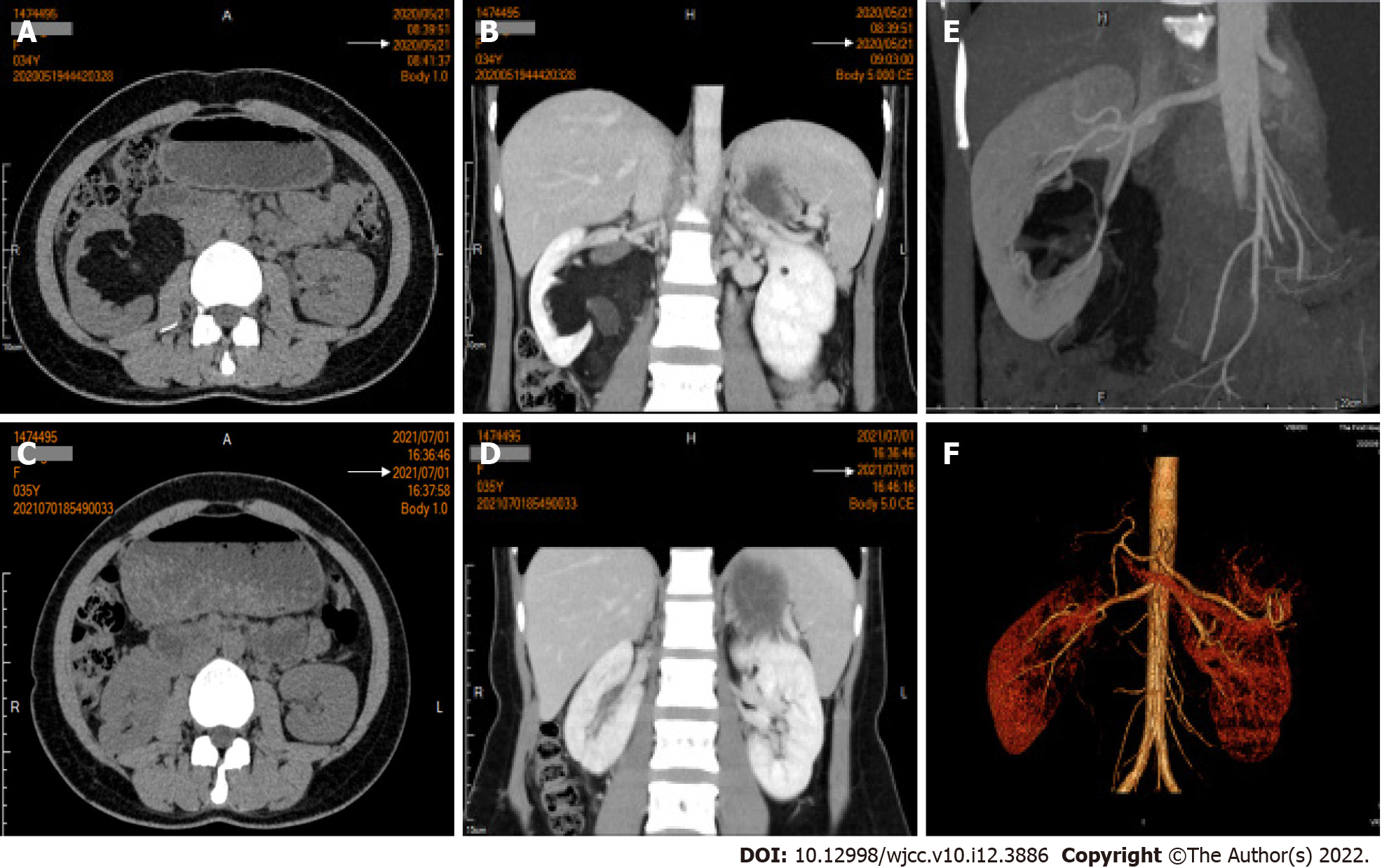
Figure 1 Computed tomography findings.
A: Plain computed tomography (CT) scan revealed a mass at the right hilum; B: Coronal section of enhanced CT image showed that the upper and lower longitude reached 9 cm; C: Plain CT scan revealed the recovery status of the right kidney at 1-year follow-up; D: Coronal section of enhanced CT image at 1-year follow-up; E: Bilateral renal CT angiography revealed the middle and lower branches of the right renal artery in the mass.
- Citation: Luo SH, Zeng QS, Chen JX, Huang B, Wang ZR, Li WJ, Yang Y, Chen LW. Successful robot-assisted partial nephrectomy for giant renal hilum angiomyolipoma through the retroperitoneal approach: A case report. World J Clin Cases 2022; 10(12): 3886-3892
- URL: https://www.wjgnet.com/2307-8960/full/v10/i12/3886.htm
- DOI: https://dx.doi.org/10.12998/wjcc.v10.i12.3886


