Copyright
©The Author(s) 2022.
World J Clin Cases. Apr 26, 2022; 10(12): 3856-3865
Published online Apr 26, 2022. doi: 10.12998/wjcc.v10.i12.3856
Published online Apr 26, 2022. doi: 10.12998/wjcc.v10.i12.3856
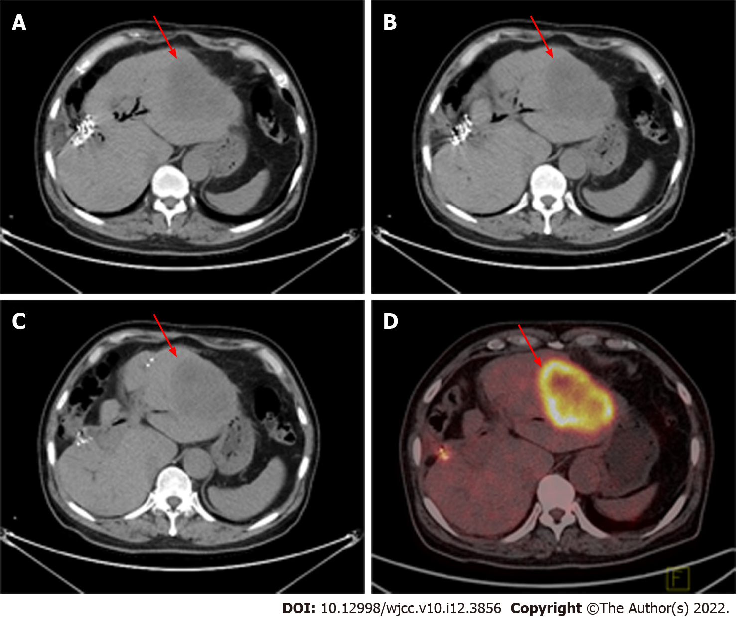
Figure 7 Abdominal computed tomography and 18F-fluorodeoxyglucose positron emission tomography/computed tomography in February 2018.
A-C: Abdominal computed tomography showed that the gallbladder had disappeared and that the liver metastasis was limited to the left liver (red arrow); D: 18F-fluorodeoxyglucose positron emission tomography/computed tomography showed that the liver metastasis was limited to the left liver (red arrow).
- Citation: Zhang B, Li S, Liu ZY, Peiris KGK, Song LF, Liu MC, Luo P, Shang D, Bi W. Successful multimodality treatment of metastatic gallbladder cancer: A case report and review of literature. World J Clin Cases 2022; 10(12): 3856-3865
- URL: https://www.wjgnet.com/2307-8960/full/v10/i12/3856.htm
- DOI: https://dx.doi.org/10.12998/wjcc.v10.i12.3856


