Copyright
©The Author(s) 2022.
World J Clin Cases. Apr 26, 2022; 10(12): 3834-3841
Published online Apr 26, 2022. doi: 10.12998/wjcc.v10.i12.3834
Published online Apr 26, 2022. doi: 10.12998/wjcc.v10.i12.3834
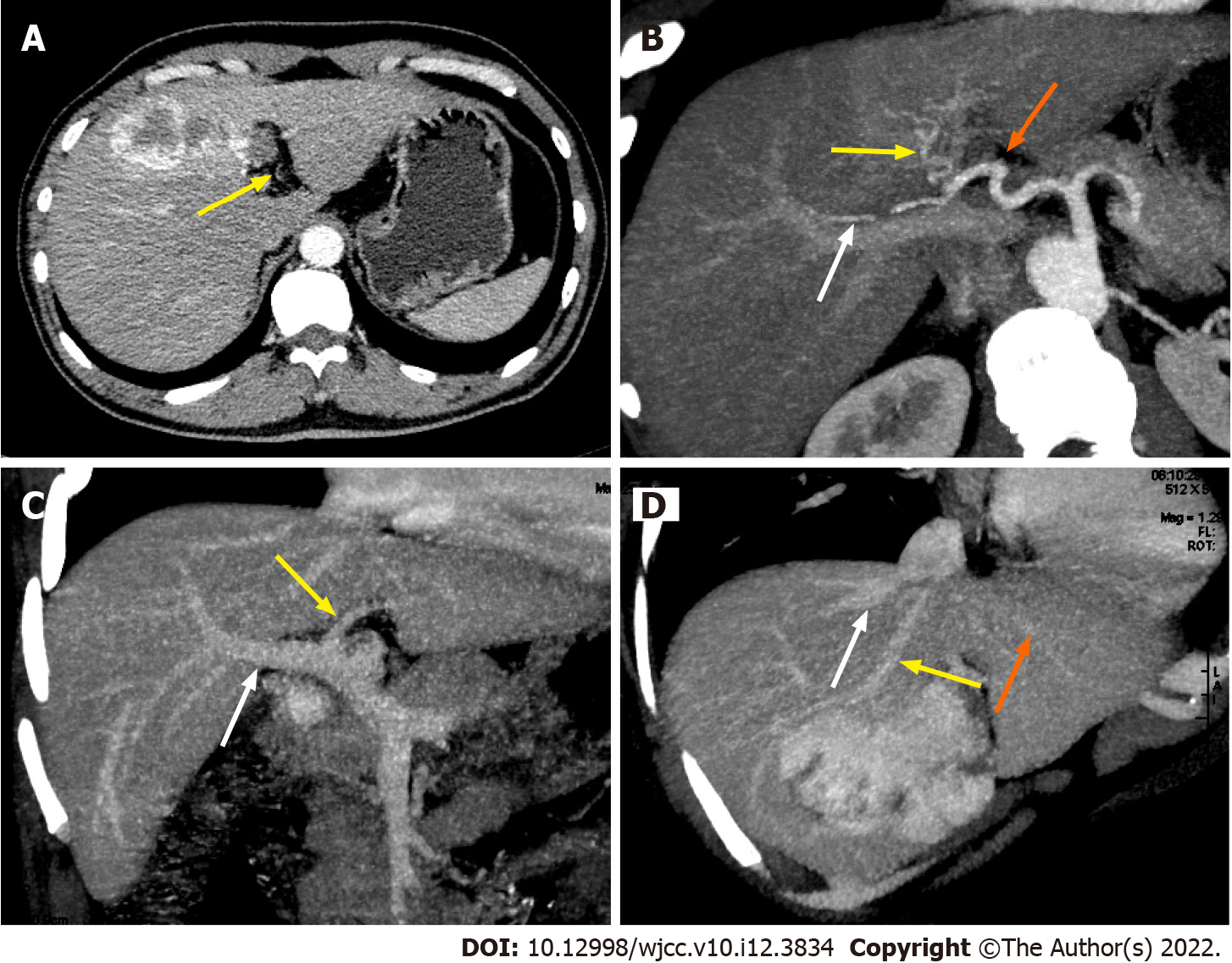
Figure 3 Preoperative contrast-enhanced abdominal computed tomography and reconstruction of the donor liver allograft vessels.
A: Yellow arrow indicate the left hepatic artery supplying the segment 2 and 3 of the liver which is not big enough to be clearly seen in reconstruction figure; B: Hepatic artery reconstruction of the liver allograft (white arrow indicate right hepatic artery supplying right lobe of the liver; yellow arrow indicate middle hepatic artery supplying segment 4 of the liver; orange arrows indicate the left hepatic artery supplying the segment 2 and 3 of the liver); C: Portal vein reconstruction of the liver allograft (white arrow indicate right portal vein; yellow arrow indicate left portal vein); D: Hepatic vein reconstruction of the live allograft (white arrow indicate right hepatic vein; yellow arrow indicate middle hepatic vein; orange arrows indicate the left hepatic vein).
- Citation: Li SX, Tang HN, Lv GY, Chen X. Pediatric living donor liver transplantation using liver allograft after ex vivo backtable resection of hemangioma: A case report. World J Clin Cases 2022; 10(12): 3834-3841
- URL: https://www.wjgnet.com/2307-8960/full/v10/i12/3834.htm
- DOI: https://dx.doi.org/10.12998/wjcc.v10.i12.3834


