Copyright
©The Author(s) 2022.
World J Clin Cases. Apr 26, 2022; 10(12): 3686-3697
Published online Apr 26, 2022. doi: 10.12998/wjcc.v10.i12.3686
Published online Apr 26, 2022. doi: 10.12998/wjcc.v10.i12.3686
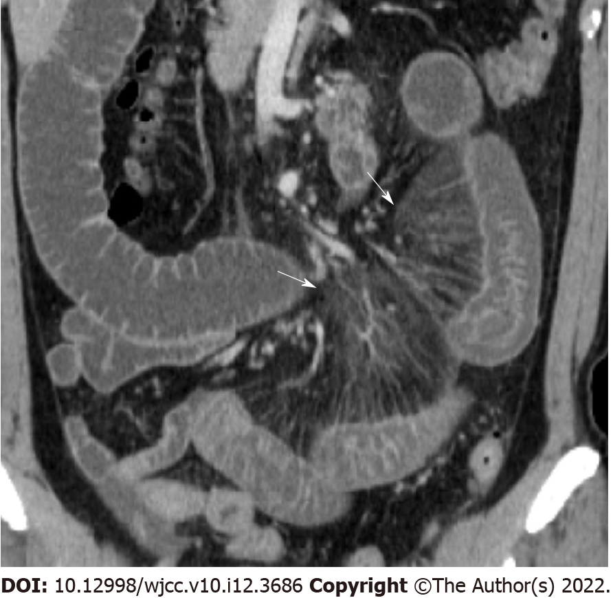
Figure 2 Comb sign.
Coronal reconstructed image shows perivascular inflammatory infiltration (arrow) that forms linear densities on the mesenteric side of the affected segments of left small bowel. Fluid distended bowel is also noted.
- Citation: Yu SJ, Heo JH, Choi EJ, Kim JH, Lee HS, Kim SY, Lim JH. Role of multidetector computed tomography in patients with acute infectious colitis. World J Clin Cases 2022; 10(12): 3686-3697
- URL: https://www.wjgnet.com/2307-8960/full/v10/i12/3686.htm
- DOI: https://dx.doi.org/10.12998/wjcc.v10.i12.3686


