Copyright
©The Author(s) 2022.
World J Clin Cases. Apr 16, 2022; 10(11): 3511-3517
Published online Apr 16, 2022. doi: 10.12998/wjcc.v10.i11.3511
Published online Apr 16, 2022. doi: 10.12998/wjcc.v10.i11.3511
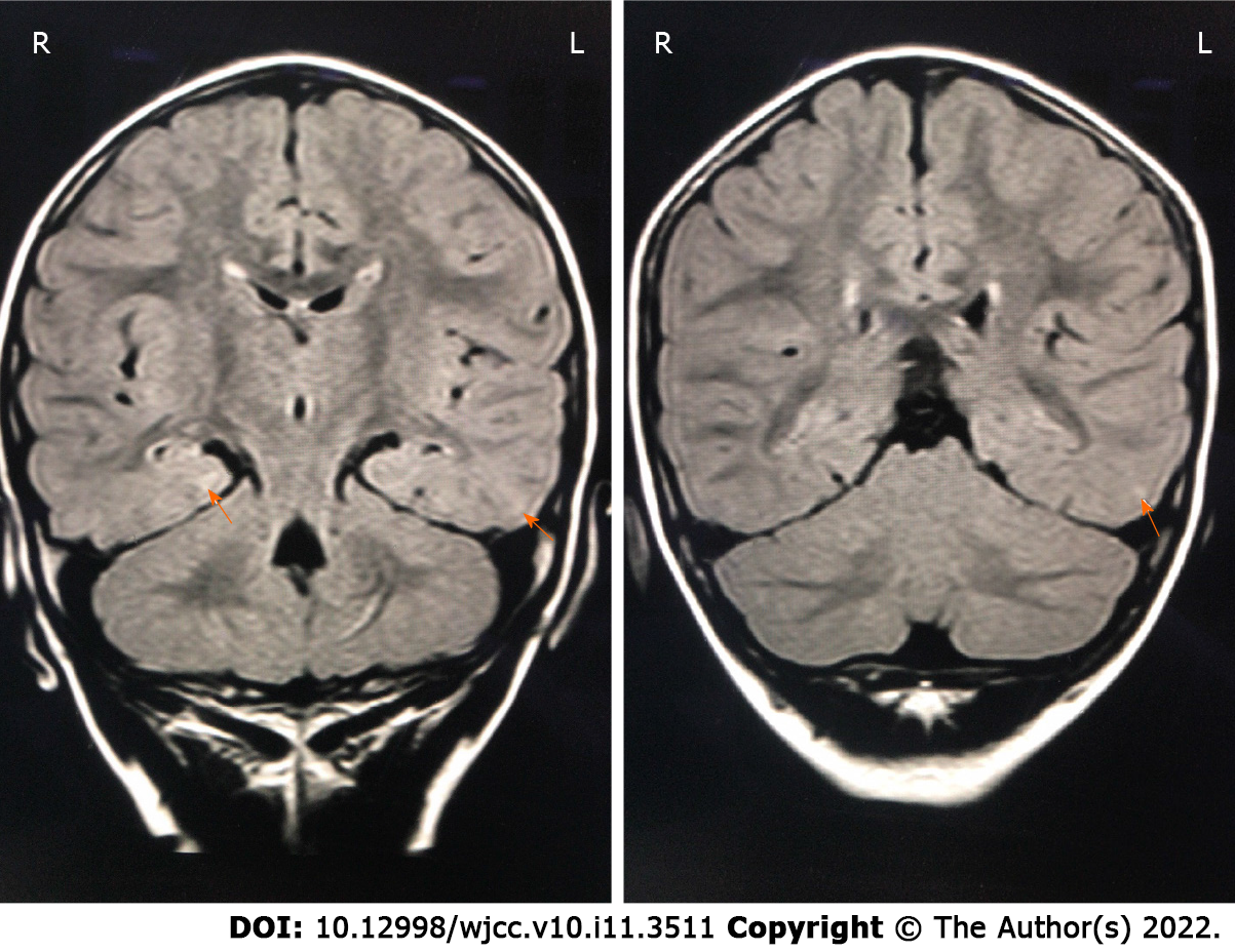
Figure 1 The patient’s cranial magnetic resonance images.
Fluid-attenuated inversion recovery showed patchy high-signal areas in the posterior part of bilateral lateral ventricle, and in the subcortical white matter of left occipital and temporal lobes (orange arrows).
- Citation: Yang H, Yu D. Young children with multidrug-resistant epilepsy and vagus nerve stimulation responding to perampanel: A case report. World J Clin Cases 2022; 10(11): 3511-3517
- URL: https://www.wjgnet.com/2307-8960/full/v10/i11/3511.htm
- DOI: https://dx.doi.org/10.12998/wjcc.v10.i11.3511


