Copyright
©The Author(s) 2022.
World J Clin Cases. Apr 16, 2022; 10(11): 3472-3477
Published online Apr 16, 2022. doi: 10.12998/wjcc.v10.i11.3472
Published online Apr 16, 2022. doi: 10.12998/wjcc.v10.i11.3472
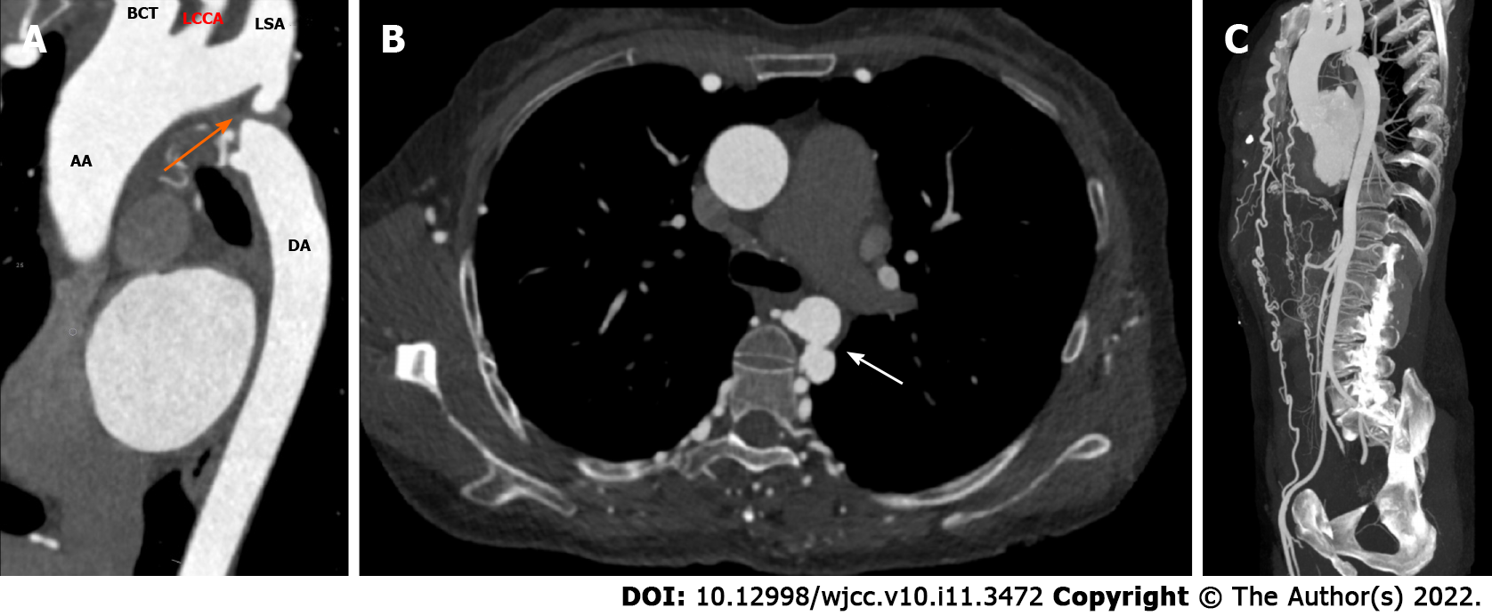
Figure 1 Type A IAA in a 54-year-old woman.
A: Reformatted oblique sagittal image of the aorta shows interruption of the aorta just distal to the subclavian artery origin (orange arrow). Note the maintenance of curvature of the ascending aorta and the extension of the interrupted aorta beyond the left subclavian artery several centimeters; B: Aneurysm formation in the descending aorta (white arrow); C: Maximum intensity projection shows a massive aorta which included two full-fledged collateral networks ensuring blood circulation to the distal aorta. AA: Ascending aorta; DA: Descending aorta; BCT: Brachiocephalic trunk; LCCA: Left common carotid artery; LSA: Left subclavian artery.
- Citation: Ren SX, Zhang Q, Li PP, Wang XD. Difference and similarity between type A interrupted aortic arch and aortic coarctation in adults: Two case reports. World J Clin Cases 2022; 10(11): 3472-3477
- URL: https://www.wjgnet.com/2307-8960/full/v10/i11/3472.htm
- DOI: https://dx.doi.org/10.12998/wjcc.v10.i11.3472


