Copyright
©The Author(s) 2022.
World J Clin Cases. Apr 16, 2022; 10(11): 3461-3471
Published online Apr 16, 2022. doi: 10.12998/wjcc.v10.i11.3461
Published online Apr 16, 2022. doi: 10.12998/wjcc.v10.i11.3461
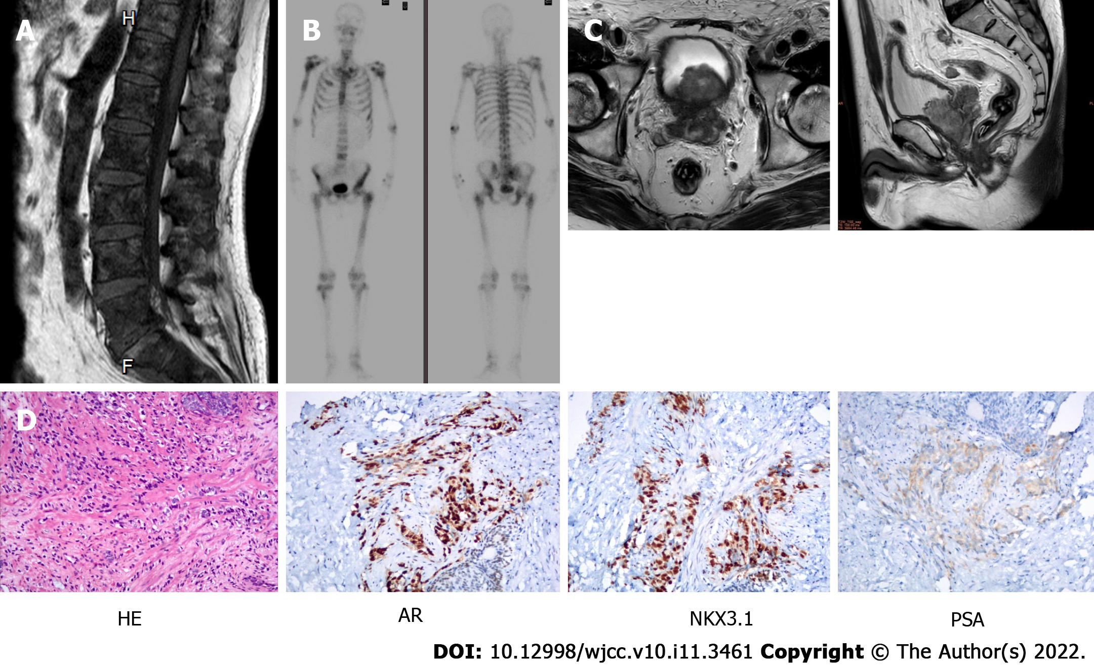
Figure 1 Radiological and immunohistochemical results showing metastatic hormone-sensitive prostate cancer.
A: Sagittal magnetic resonance imaging (MRI) showed multiple lumbar vertebrate metastases; B: Bone scanning displayed the high metabolic activity of bone lesions; C: Cross (left) and sagittal (right) sections of MRI scanning (T2WI) displayed invasion of the prostate malignant lesion into the bladder wall as well as the seminal vesicle; D: Hematoxylin-eosinstaining and immunohistochemistry results for androgen receptor, NKX3.1, and periodic Schiff Acid staining. Original magnification: 100 ×; scale bar: 100 μm. HE: Hematoxylin-eosin; AR: Androgen receptor; PSA: Periodic Schiff Acid.
- Citation: Yuan F, Liu N, Yang MZ, Zhang XT, Luo H, Zhou H. Circulating tumor DNA genomic profiling reveals the complicated olaparib-resistance mechanism in prostate cancer salvage therapy: A case report. World J Clin Cases 2022; 10(11): 3461-3471
- URL: https://www.wjgnet.com/2307-8960/full/v10/i11/3461.htm
- DOI: https://dx.doi.org/10.12998/wjcc.v10.i11.3461


