Copyright
©The Author(s) 2022.
World J Clin Cases. Apr 16, 2022; 10(11): 3369-3378
Published online Apr 16, 2022. doi: 10.12998/wjcc.v10.i11.3369
Published online Apr 16, 2022. doi: 10.12998/wjcc.v10.i11.3369
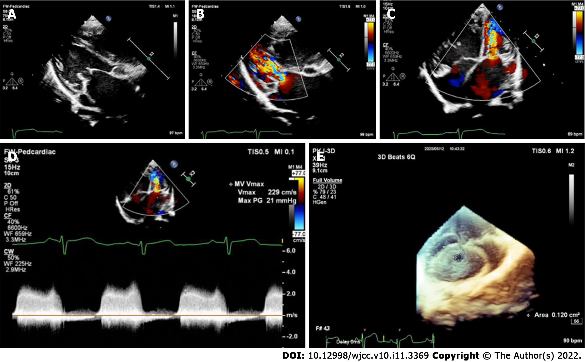
Figure 1 A case of Annulo-Leaflet mitral ring.
A: Two-dimensional ultrasonography at the parasternal left ventricular long-axis view revealed thickened mitral valve and a mitral ring adhered to the anterior and posterior leaflets of the mitral valve, restricting the opening of the valve leaflets and causing mitral valve inflow tract obstruction; B: Color Doppler ultrasonography at the parasternal left ventricular long-axis view revealed that blood flow velocity increased at the mitral annulus, and the forward flow velocity of the mitral valve was increased; C: Color Doppler ultrasonography at the apical four-chamber view revealed that blood flow velocity increased at the mitral annulus, and the forward flow velocity of the mitral valve was increased; D: The forward flow velocity of the mitral valve was increased significantly, at 2.29 m/s; E: Real-time three-dimensional echocardiography showed a narrow mitral valve orifice.
- Citation: Li YD, Meng H, Pang KJ, Li MZ, Xu N, Wang H, Li SJ, Yan J. Echocardiography in the diagnosis of Shone’s complex and analysis of the causes for missed diagnosis and misdiagnosis. World J Clin Cases 2022; 10(11): 3369-3378
- URL: https://www.wjgnet.com/2307-8960/full/v10/i11/3369.htm
- DOI: https://dx.doi.org/10.12998/wjcc.v10.i11.3369


