Copyright
©The Author(s) 2022.
World J Clin Cases. Apr 6, 2022; 10(10): 3306-3312
Published online Apr 6, 2022. doi: 10.12998/wjcc.v10.i10.3306
Published online Apr 6, 2022. doi: 10.12998/wjcc.v10.i10.3306
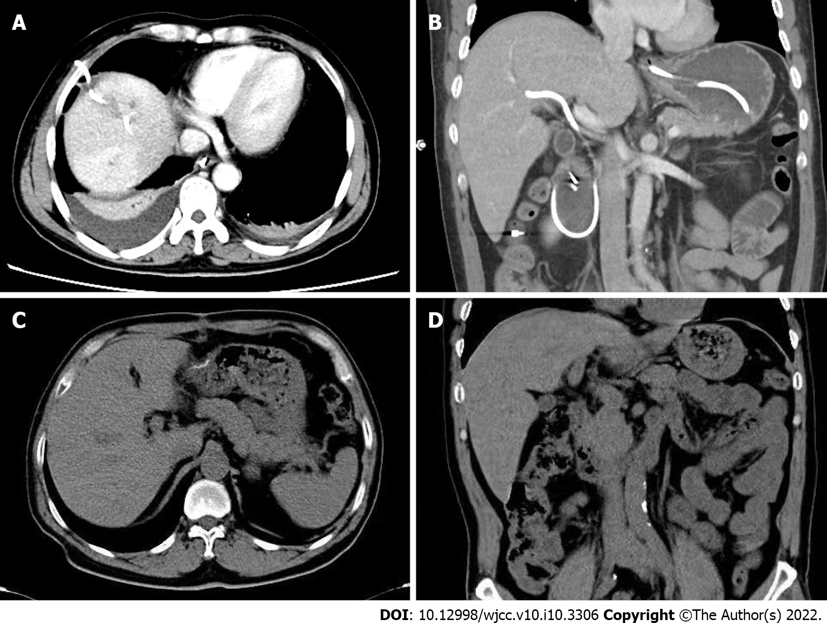
Figure 4 Abdominal computed tomography scan.
A and B: Abdominal computed tomography scan was performed on postoperative day 10. Liver abscess was absorbed by the ultrasound-guided percutaneous catheter drainage (A); Endoscopic nasobiliary drainage tube was placed in the right anterior bile duct without displacement (B); C and D: Abdominal computed tomography scan was performed 1 mo later. There was no intrahepatic mass found (C); There was no expansion of the bile duct inside and outside the liver (D).
- Citation: Zou H, Wen Y, Pang Y, Zhang H, Zhang L, Tang LJ, Wu H. Endoscopic-catheter-directed infusion of diluted (-)-noradrenaline for atypical hemobilia caused by liver abscess: A case report. World J Clin Cases 2022; 10(10): 3306-3312
- URL: https://www.wjgnet.com/2307-8960/full/v10/i10/3306.htm
- DOI: https://dx.doi.org/10.12998/wjcc.v10.i10.3306


