Copyright
©The Author(s) 2022.
World J Clin Cases. Apr 6, 2022; 10(10): 3251-3260
Published online Apr 6, 2022. doi: 10.12998/wjcc.v10.i10.3251
Published online Apr 6, 2022. doi: 10.12998/wjcc.v10.i10.3251
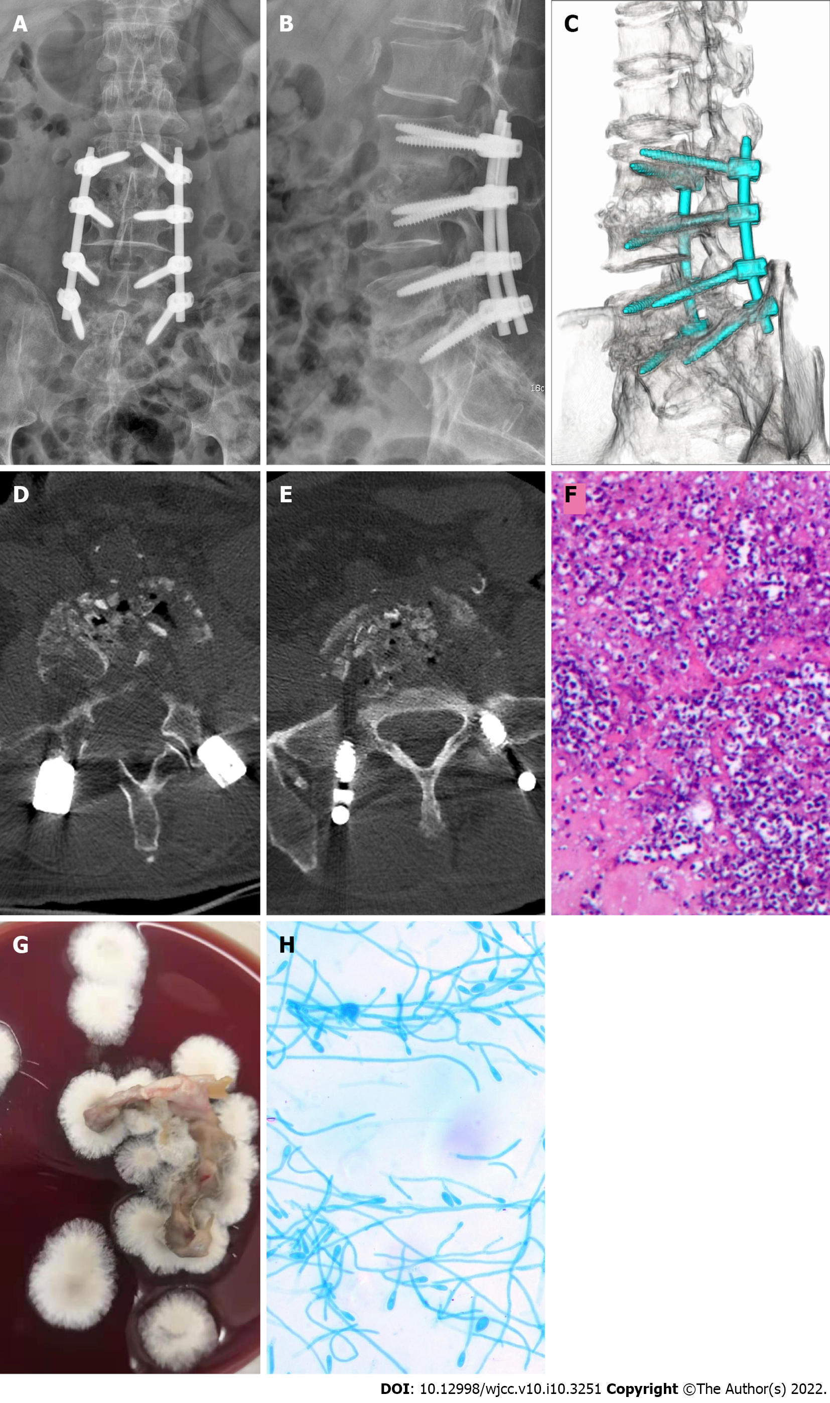
Figure 2 Medical imaging, pathology and microbiology examination after surgery.
A and B: X-ray image showing that the lumbar internal fixation device was in a good position; C-E: Three-dimensional computed tomography image showing sufficient bone grafting in the L3/L4 and L5/S1 intervertebral spaces; F: Pathological examination results showing a large number of inflammatory cells in the tissues examined, and the staining revealed PAS (+), and acid resistance (-); G: Lesion tissue culture day 7 (blood agar medium, 30 °C, 7 d) showed that colonies were cashmere-like and the back was gray-black; H: Under the microscope (lactic acid phenol cotton blue staining, × 400), most of the hyphae were irregularly branched, producing round or oval lateral and terminal conidia.
- Citation: Shi XW, Li ST, Lou JP, Xu B, Wang J, Wang X, Liu H, Li SK, Zhen P, Zhang T. Scedosporium apiospermum infection of the lumbar vertebrae: A case report. World J Clin Cases 2022; 10(10): 3251-3260
- URL: https://www.wjgnet.com/2307-8960/full/v10/i10/3251.htm
- DOI: https://dx.doi.org/10.12998/wjcc.v10.i10.3251


