Copyright
©The Author(s) 2022.
World J Clin Cases. Apr 6, 2022; 10(10): 3170-3177
Published online Apr 6, 2022. doi: 10.12998/wjcc.v10.i10.3170
Published online Apr 6, 2022. doi: 10.12998/wjcc.v10.i10.3170
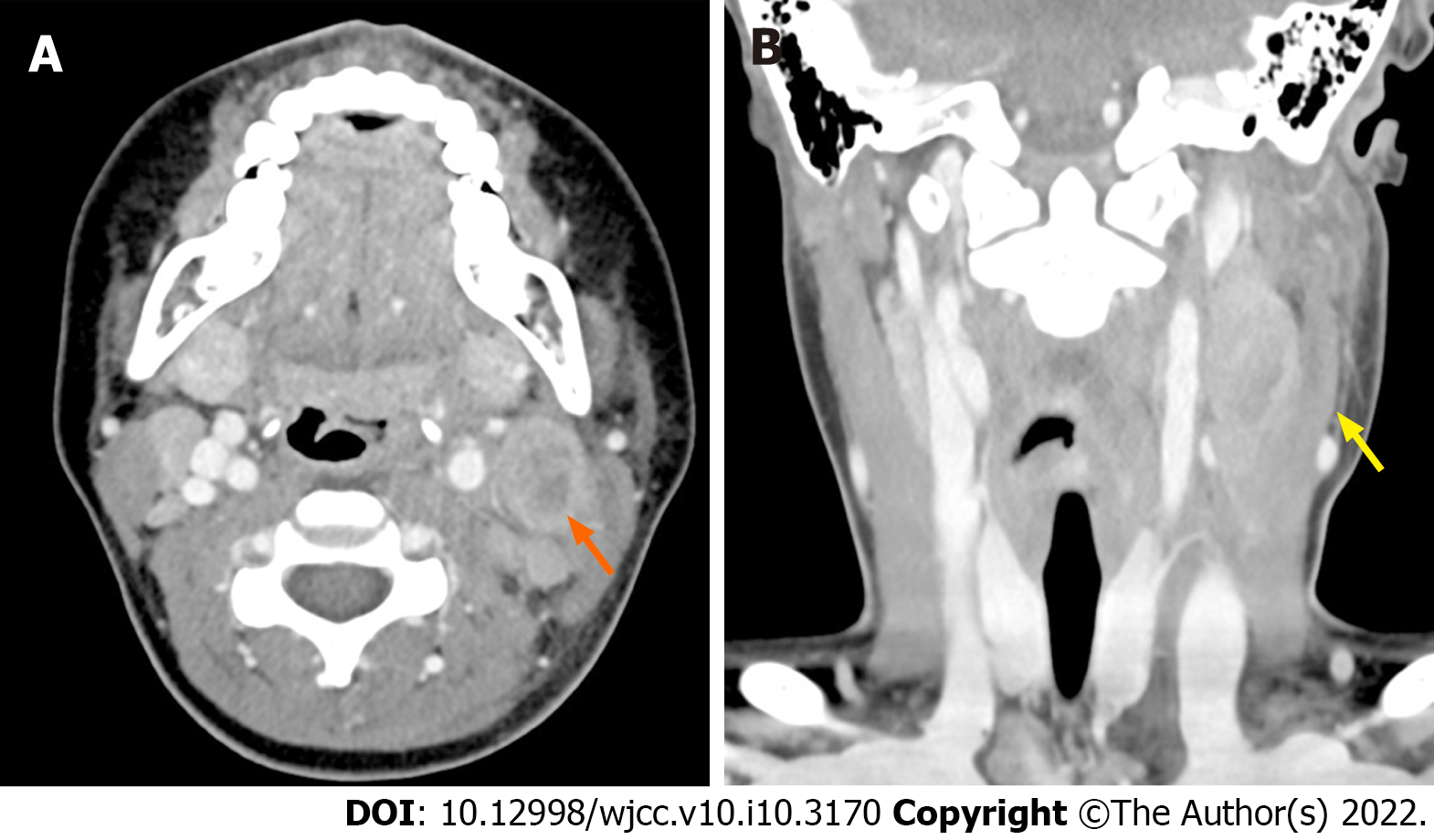
Figure 1 Neck computed tomography horizontal (A) and coronal (B) views showing perilesional infiltration (yellow arrow) and necrosis (orange arrow) as acute infectious lymphadenitis.
Neck computed tomography reveals a 2.5 cm × 2.0 cm × 3.2 cm-sized acute infectious lymphadenitis with perilesional infiltration and necrosis.
- Citation: Kim N, Choi YJ, Na JY, Oh JW. Lymph-node-first presentation of Kawasaki disease in a 12-year-old girl with cervical lymphadenitis caused by Mycoplasma pneumoniae: A case report. World J Clin Cases 2022; 10(10): 3170-3177
- URL: https://www.wjgnet.com/2307-8960/full/v10/i10/3170.htm
- DOI: https://dx.doi.org/10.12998/wjcc.v10.i10.3170


