Copyright
©The Author(s) 2022.
World J Clin Cases. Jan 7, 2022; 10(1): 388-396
Published online Jan 7, 2022. doi: 10.12998/wjcc.v10.i1.388
Published online Jan 7, 2022. doi: 10.12998/wjcc.v10.i1.388
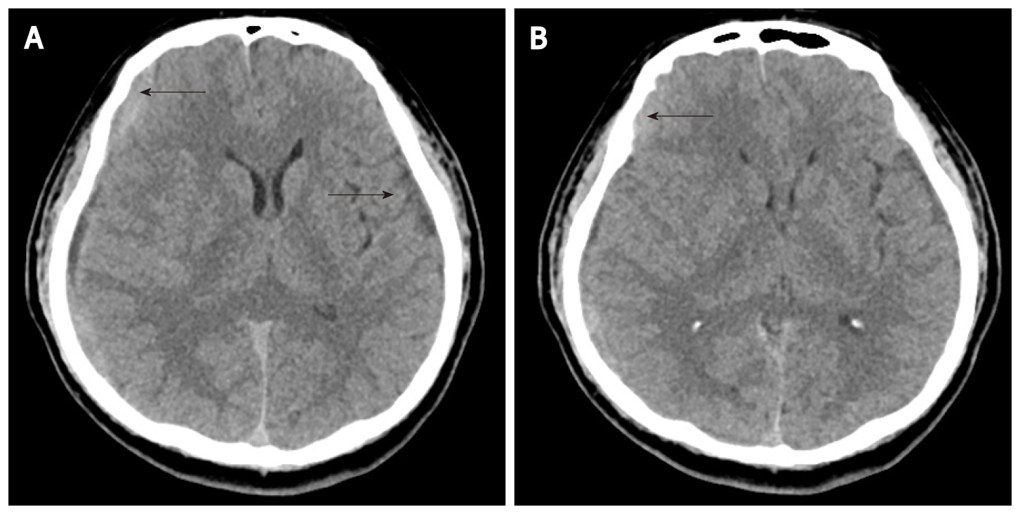
Figure 5 Follow-up axial computerized tomographic scanning.
A: Reduced attenuation of subdural hematoma (SDH) along the left frontoparietotemporal cerebral convexities; B: Reduced amount of SDH along the right frontoparietotemporal cerebral convexities. Black arrow: SDH.
- Citation: Choi SH, Lee YY, Kim WJ. Epidural blood patch for spontaneous intracranial hypotension with subdural hematoma: A case report and review of literature. World J Clin Cases 2022; 10(1): 388-396
- URL: https://www.wjgnet.com/2307-8960/full/v10/i1/388.htm
- DOI: https://dx.doi.org/10.12998/wjcc.v10.i1.388


