Copyright
©The Author(s) 2022.
World J Clin Cases. Jan 7, 2022; 10(1): 331-337
Published online Jan 7, 2022. doi: 10.12998/wjcc.v10.i1.331
Published online Jan 7, 2022. doi: 10.12998/wjcc.v10.i1.331
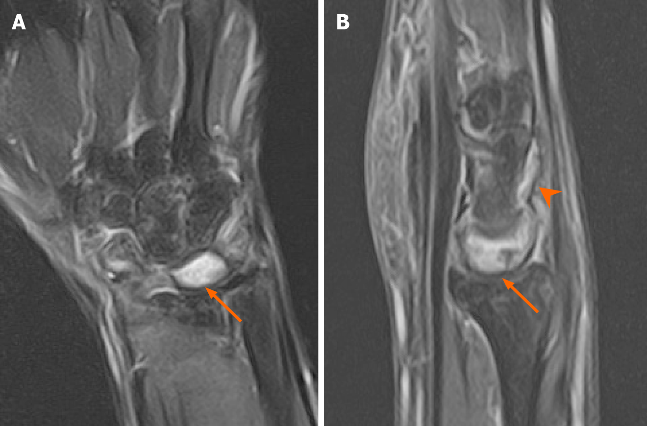
Figure 3 Two months postoperative magnetic resonance imaging showed residual bone edema of the lunate.
A: Magnetic resonance imaging (MRI) in T2W coronal Fat saturation (FS) showed residual bone marrow edema in scaphoid, triquetrum and particularly, the lunate (arrow). This finding might be attributed to the post-traumatic or postoperative change; B: MRI in T2W sagittal FS showed lunate bone marrow edema and effusion in the intercarpal joints (arrowhead).
- Citation: Li LY, Lin CJ, Ko CY. Lunate dislocation with avulsed triquetral fracture: A case report. World J Clin Cases 2022; 10(1): 331-337
- URL: https://www.wjgnet.com/2307-8960/full/v10/i1/331.htm
- DOI: https://dx.doi.org/10.12998/wjcc.v10.i1.331


