Copyright
©The Author(s) 2022.
World J Clin Cases. Jan 7, 2022; 10(1): 331-337
Published online Jan 7, 2022. doi: 10.12998/wjcc.v10.i1.331
Published online Jan 7, 2022. doi: 10.12998/wjcc.v10.i1.331
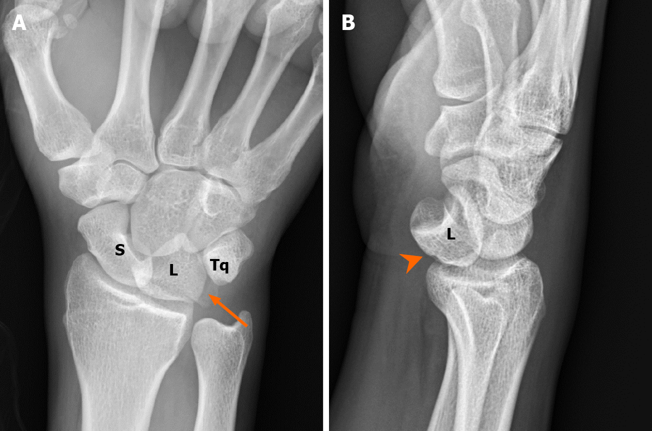
Figure 1 A 37-year-old man with multiple traumas.
Radiological examinations revealed acute lunate dislocation with triquetral fracture. A: Plain radiograph in the emergency department showed triangular appearance of the lunate (piece of pie) with triquetral fracture. Note the avulsion of the triquetrum (arrow) due to failure of the lunotriquetral ligaments; B: Lateral view of the radiograph showed volar dislocation of the lunate bone (arrowhead). S: Scaphoid, L: Lunate, Tq: Triquetrum.
- Citation: Li LY, Lin CJ, Ko CY. Lunate dislocation with avulsed triquetral fracture: A case report. World J Clin Cases 2022; 10(1): 331-337
- URL: https://www.wjgnet.com/2307-8960/full/v10/i1/331.htm
- DOI: https://dx.doi.org/10.12998/wjcc.v10.i1.331


