Copyright
©The Author(s) 2022.
World J Clin Cases. Jan 7, 2022; 10(1): 323-330
Published online Jan 7, 2022. doi: 10.12998/wjcc.v10.i1.323
Published online Jan 7, 2022. doi: 10.12998/wjcc.v10.i1.323
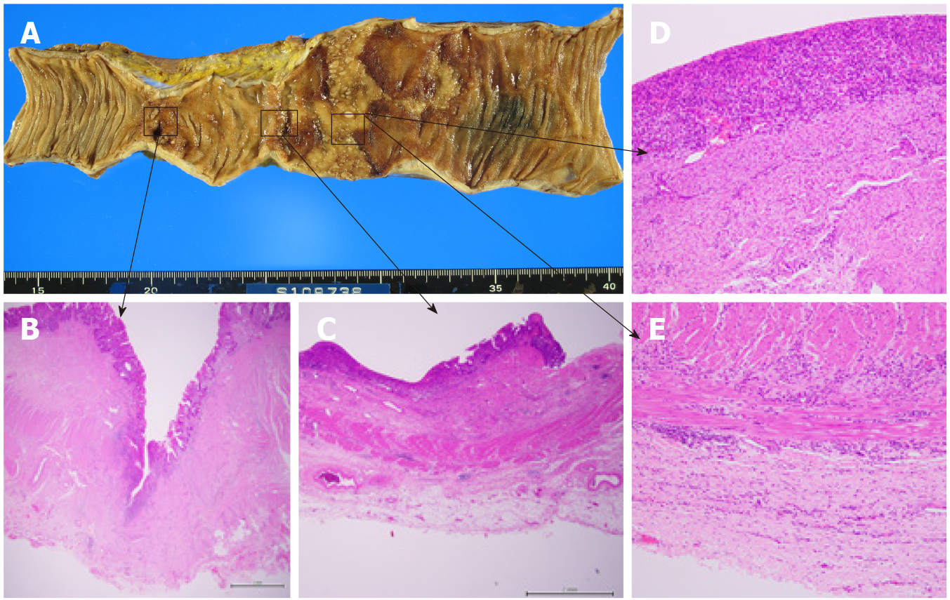
Figure 3 Macroscopic and microscopic findings.
A: Findings revealed a deep ulcer with stenosis and a shallow ulcer in continuous erosion, along with normal mucosa marked with Indian ink on the oral side; B: Pathological findings revealed a deep-mining ulcer (to the muscularis propria) with collagen hyperplasia; C: A shallow ulcer with epithelial loss; D: An abscess, with a shallow ulcer with chronic inflammatory cell infiltration from the mucosa to the subserosa; E: A shallow ulcer with chronic inflammatory cell infiltration from the mucosa to the subserosa.
- Citation: Yasuda T, Sakurazawa N, Kuge K, Omori J, Arai H, Kakinuma D, Watanabe M, Suzuki H, Iwakiri K, Yoshida H. Protein-losing enteropathy caused by a jejunal ulcer after an internal hernia in Petersen's space: A case report. World J Clin Cases 2022; 10(1): 323-330
- URL: https://www.wjgnet.com/2307-8960/full/v10/i1/323.htm
- DOI: https://dx.doi.org/10.12998/wjcc.v10.i1.323


