Copyright
©The Author(s) 2022.
World J Clin Cases. Jan 7, 2022; 10(1): 275-282
Published online Jan 7, 2022. doi: 10.12998/wjcc.v10.i1.275
Published online Jan 7, 2022. doi: 10.12998/wjcc.v10.i1.275
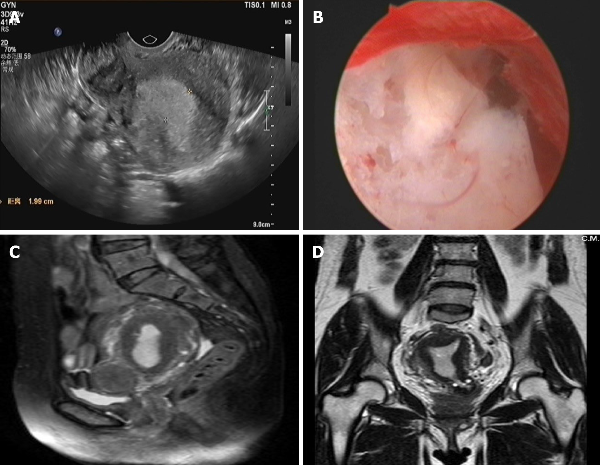
Figure 1 Imaging findings of the patient before the fourth operation.
A: Transvaginal ultrasonography: The endometrium was thickened (approximately 2.0 cm in total). Several hypoechoic masses were observed in the uterine area. There was no obvious space-occupying lesion in bilateral adnexa; B: Hysteroscopy: the endometrium was extensively thickened, the texture was fragile; C, D: Pelvic MR: The endometrium was thickened (approximately 1.6 cm in total), and the signal in the right corner of the uterine cavity was not uniform.
- Citation: Wang J, Yang Q, Zhang NN, Wang DD. Recurrent postmenopausal bleeding - just endometrial disease or ovarian sex cord-stromal tumor? A case report. World J Clin Cases 2022; 10(1): 275-282
- URL: https://www.wjgnet.com/2307-8960/full/v10/i1/275.htm
- DOI: https://dx.doi.org/10.12998/wjcc.v10.i1.275


