Copyright
©The Author(s) 2022.
World J Clin Cases. Jan 7, 2022; 10(1): 268-274
Published online Jan 7, 2022. doi: 10.12998/wjcc.v10.i1.268
Published online Jan 7, 2022. doi: 10.12998/wjcc.v10.i1.268
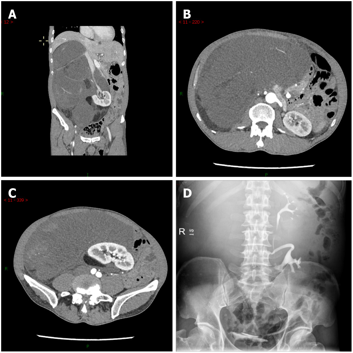
Figure 1 Imaging findings before treatment.
A and C: Computed tomography angiography showed a massive lipoma-like mass extending from the sub-hepatic space to the pelvic cavity, with multiple organs dislocated; B and D: Intravenous pyelography with radiocontrast agent confirmed the displacement of the right kidney to the left lower quadrant and its excretion function was good.
- Citation: Ye MS, Wu HK, Qin XZ, Luo F, Li Z. Hyper-accuracy three-dimensional reconstruction as a tool for better planning of retroperitoneal liposarcoma resection: A case report. World J Clin Cases 2022; 10(1): 268-274
- URL: https://www.wjgnet.com/2307-8960/full/v10/i1/268.htm
- DOI: https://dx.doi.org/10.12998/wjcc.v10.i1.268


