
















©The Author(s) 2022.
World J Clin Cases. Jan 7, 2022; 10(1): 217-226
Published online Jan 7, 2022. doi: 10.12998/wjcc.v10.i1.217
Published online Jan 7, 2022. doi: 10.12998/wjcc.v10.i1.217
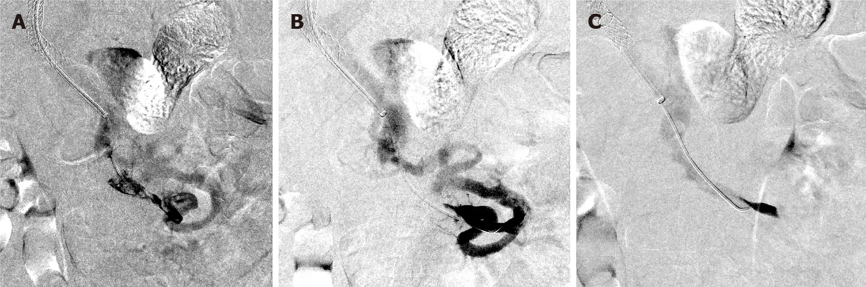
Figure 2 Laboratory tests, imaging, and treatment modalities.
A and B: Selective superior mesenteric venogram after transjugular intrahepatic portosystemic shunt creation demonstrates a filling defect representing thrombus in the superior mesenteric venous (SMV), hepatofugal flow in a peripheral SMV branch, and a venovenous collateral decompressing the small bowel outflow into the main SMV trunk; C: Superior mesenteric venogram after aspiration and mechanical thrombectomy demonstrates reestablished flow in the previously occluded SMV, nonperfusion for the venovenous collateral, and hepatopetal flow in the peripheral SMV branches.
- Citation: Alnahhal K, Toskich BB, Nussbaum S, Li Z, Erben Y, Hakaim AG, Farres H. Superior mesenteric venous thrombosis: Endovascular management and outcomes. World J Clin Cases 2022; 10(1): 217-226
- URL: https://www.wjgnet.com/2307-8960/full/v10/i1/217.htm
- DOI: https://dx.doi.org/10.12998/wjcc.v10.i1.217