Copyright
©2013 Baishideng Publishing Group Co.
World J Clin Cases. Jul 16, 2013; 1(4): 152-154
Published online Jul 16, 2013. doi: 10.12998/wjcc.v1.i4.152
Published online Jul 16, 2013. doi: 10.12998/wjcc.v1.i4.152
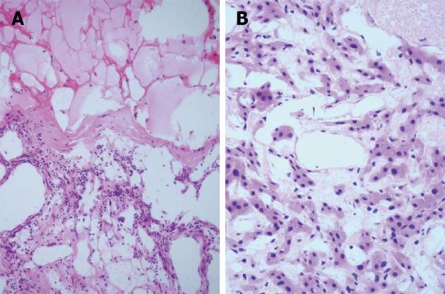
Figure 3 Histopathological findings.
A: Dilated lymphatic cavities accompanied with partial infarction. Lymph cells were seen lining the wall of the cyst, hematoxylin and eosin (HE), × 100; B: Simple squamous epithelia and eosinophilic hepatic cells (HE, × 200).
- Citation: Zhang YZ, Ye YS, Tian L, Li W. Rare case of a solitary huge hepatic cystic lymphangioma. World J Clin Cases 2013; 1(4): 152-154
- URL: https://www.wjgnet.com/2307-8960/full/v1/i4/152.htm
- DOI: https://dx.doi.org/10.12998/wjcc.v1.i4.152


