Copyright
©The Author(s) 2021.
World J Clin Cases. Feb 6, 2021; 9(4): 830-837
Published online Feb 6, 2021. doi: 10.12998/wjcc.v9.i4.830
Published online Feb 6, 2021. doi: 10.12998/wjcc.v9.i4.830
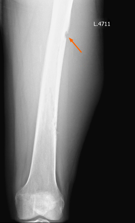
Figure 1 An X-Ray of the left thigh.
Orange arrow indicates start of destruction of the femur.
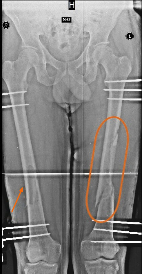
Figure 2 An X-Ray of both thighs with external AO fixation (preventive on the right).
Orange arrow shows sequester, orange circle indicates osteosinthesis after fracture with displacement.
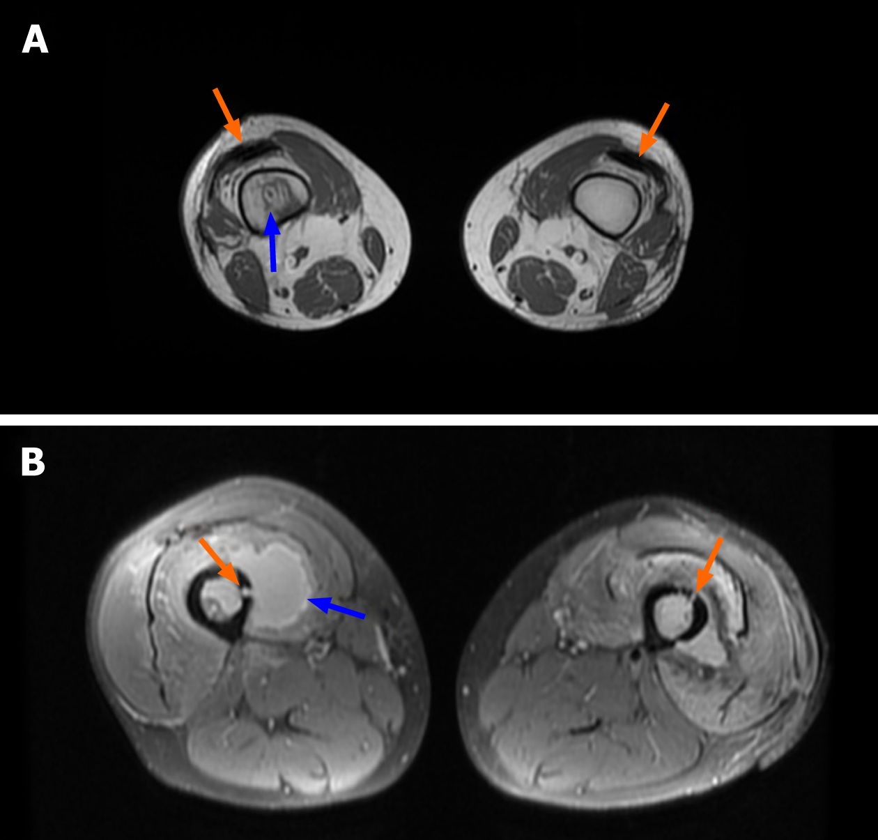
Figure 3 MRI scan of both thighs, axial section.
A: Orange arrows indicate air inserts in interfascial spaces. Blue arrow indicates an abscess in the intramedullary space in the right femur. B: Fistulas are marked with orange arrows merging with the intramedullary space, present bone destruction. The blue arrow indicates a clearly visible abscess around the femur.
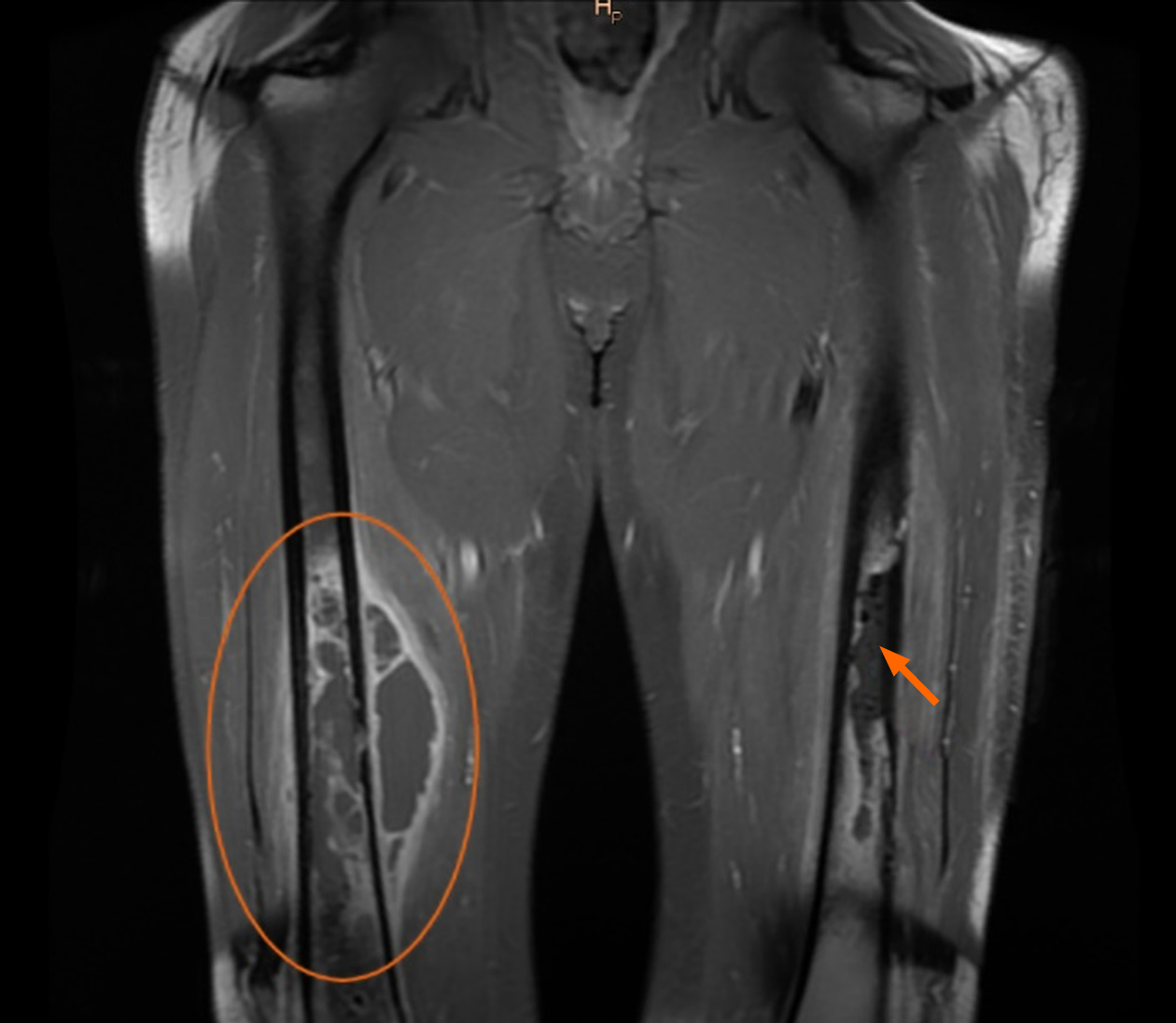
Figure 4 MRI scan of both thighs, frontal section.
The orange circle shows the abscess near the femur and in intramedullary space. The orange arrow indicates the destructive processes taking place in the left femur. Also, the photo shows the longitudinal air inserts in the interfascial space.
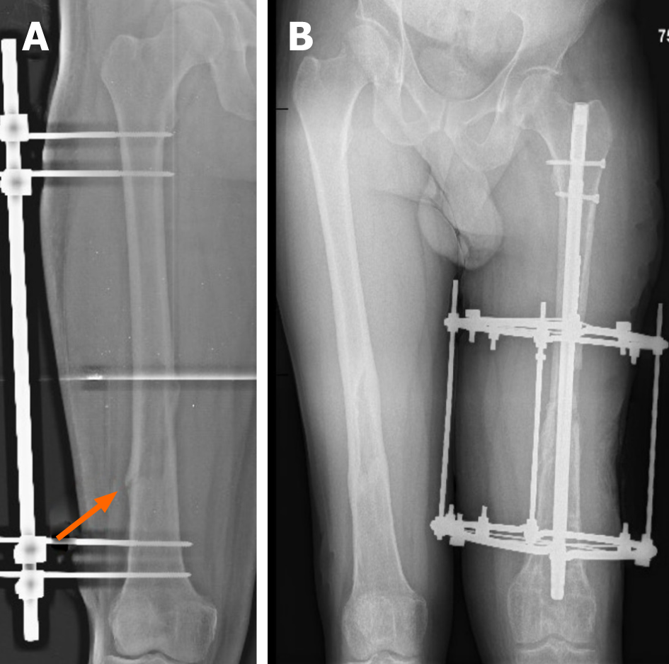
Figure 5 An X-Ray of both femurs after occurrence of pathological fractures.
A: Pathological fracture occurred in the right femur, despite previous external AO fixation. Orange arrow indicates the location of the fracture B: An X-Ray of both thighs. Right femur completely healed (after 6 mo of treatment). On the left: External fixation with Ilizarov’s apparatus, intramedullary osteosynthesis with silver-plated intramedullary nail. An X-Ray shows a significant shortening of the left limb.
- Citation: Daunaraite K, Uvarovas V, Ulevicius D, Sveikata T, Petryla G, Kurtinaitis J, Satkauskas I. Reciprocal hematogenous osteomyelitis of the femurs caused by Anaerococcus prevotii: A case report. World J Clin Cases 2021; 9(4): 830-837
- URL: https://www.wjgnet.com/2307-8960/full/v9/i4/830.htm
- DOI: https://dx.doi.org/10.12998/wjcc.v9.i4.830


