Copyright
©The Author(s) 2021.
World J Clin Cases. Sep 26, 2021; 9(27): 8260-8267
Published online Sep 26, 2021. doi: 10.12998/wjcc.v9.i27.8260
Published online Sep 26, 2021. doi: 10.12998/wjcc.v9.i27.8260
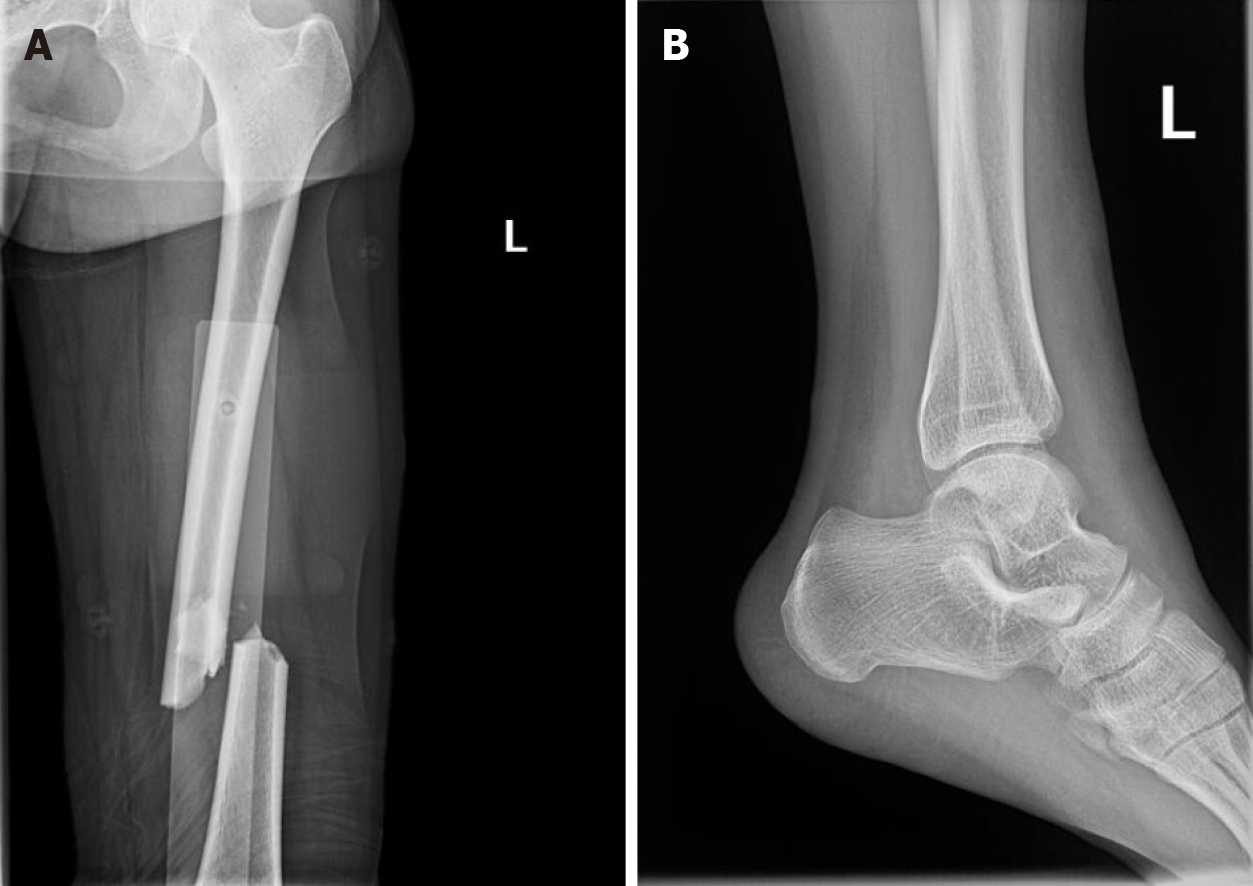
Figure 1 X-ray examination.
A: A fracture of the middle and lower part of the left femur; B: A fracture of the base of the left fifth metatarsal bone.
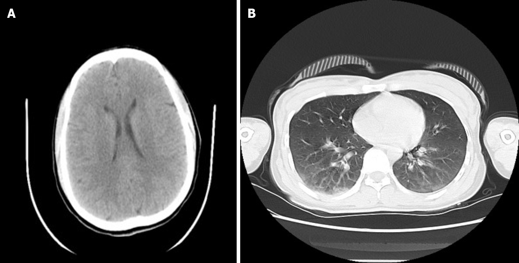
Figure 2 Computed tomography examination.
A: The results of computed tomography (CT) showed that there was no obvious abnormality in the skull; B: The results of CT showed that there was no obvious abnormality and only minor contusionin in the lung.
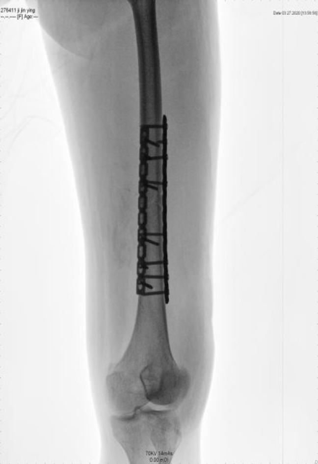
Figure 3
Open reduction and internal fixation on the femur.
- Citation: Yang J, Cui ZN, Dong JN, Lin WB, Jin JT, Tang XJ, Guo XB, Cui SB, Sun M, Ji CC. Early acute fat embolism syndrome caused by femoral fracture: A case report. World J Clin Cases 2021; 9(27): 8260-8267
- URL: https://www.wjgnet.com/2307-8960/full/v9/i27/8260.htm
- DOI: https://dx.doi.org/10.12998/wjcc.v9.i27.8260


