Copyright
©The Author(s) 2021.
World J Clin Cases. May 26, 2021; 9(15): 3787-3795
Published online May 26, 2021. doi: 10.12998/wjcc.v9.i15.3787
Published online May 26, 2021. doi: 10.12998/wjcc.v9.i15.3787
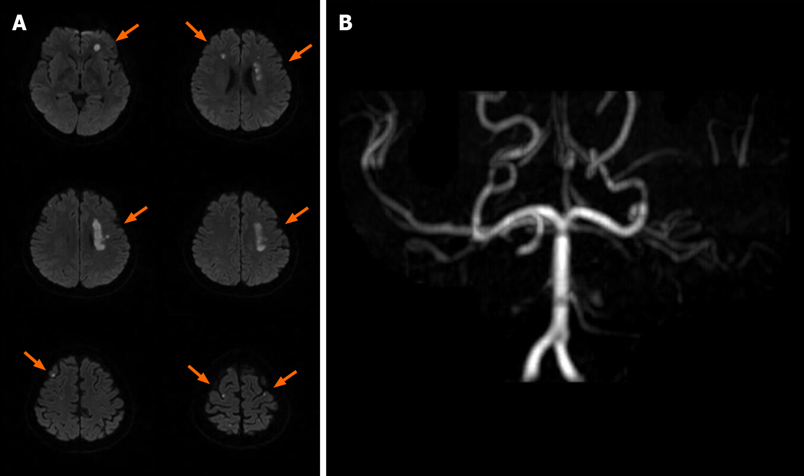
Figure 1 Brain magnetic resonance imaging of the first stroke before admission.
A: The diffusion weighted imaging shows left corona radiata, bilateral frontal lobe, and parietal lobe infarction (orange arrow); B: The magnetic resonance angiography presents bilateral internal carotid artery occlusion.
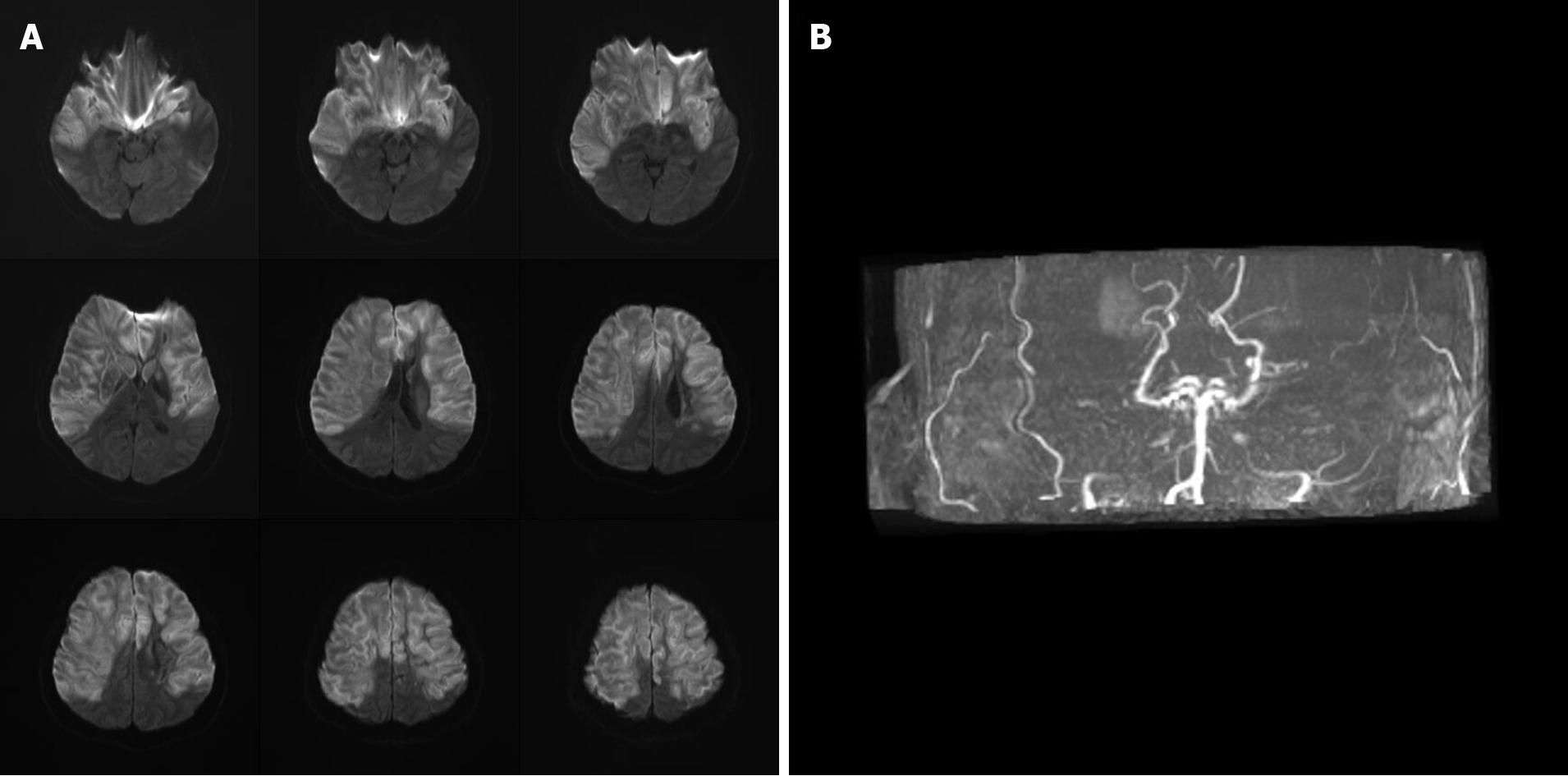
Figure 2 Brain magnetic resonance imaging of the second stroke after diabetic ketoacidosis.
A: The diffusion weighted imaging shows: (1) Acute infarction over the bilateral middle cerebral artery and bilateral anterior cerebral artery territory with brain swelling of infarction lesions and midline shift; and (2) Occlusion of bilateral internal carotid artery; B: The magnetic resonance angiography also presents bilateral internal carotid artery occlusion.
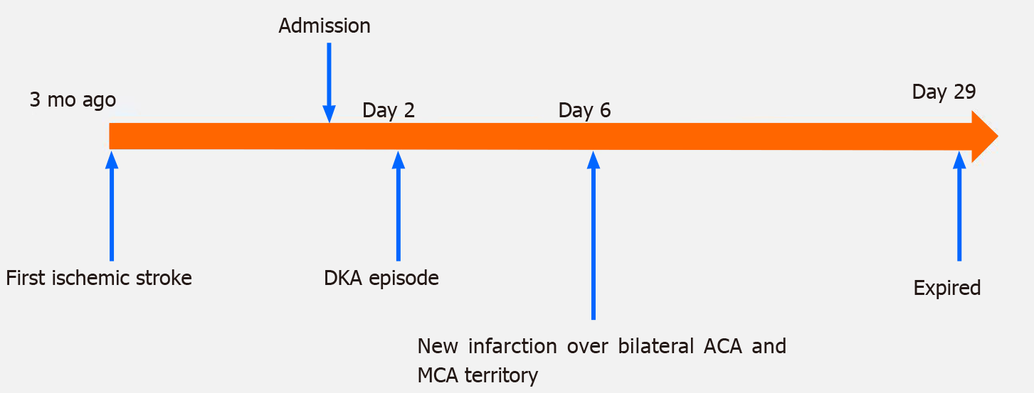
Figure 3 Timeline of this patient.
DKA: Diabetic ketoacidosis; ACA: Anterior cerebral artery; MCA: Middle cerebral artery.
- Citation: Chen YC, Tsai SJ. Bilateral cerebral infarction in diabetic ketoacidosis and bilateral internal carotid artery occlusion: A case report and review of literature. World J Clin Cases 2021; 9(15): 3787-3795
- URL: https://www.wjgnet.com/2307-8960/full/v9/i15/3787.htm
- DOI: https://dx.doi.org/10.12998/wjcc.v9.i15.3787


