Copyright
©The Author(s) 2020.
World J Clin Cases. Dec 26, 2020; 8(24): 6337-6345
Published online Dec 26, 2020. doi: 10.12998/wjcc.v8.i24.6337
Published online Dec 26, 2020. doi: 10.12998/wjcc.v8.i24.6337
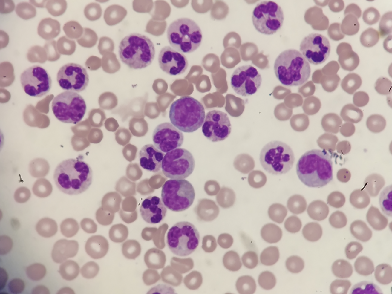
Figure 1 Histology of blood.
Peripheral blood smear (Wright-Giemsa staining, × 500).
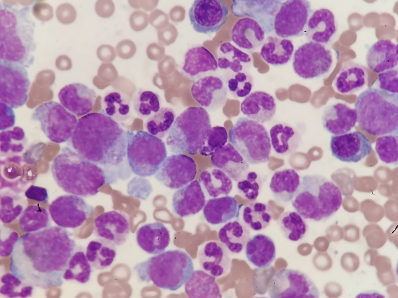
Figure 2 Histology of bone marrow biopsy.
Bone marrow aspirate smear showing granulocytic proliferation, many mature neutrophils with toxic changes, and no increased blasts (Wright-Giemsa staining, × 500).
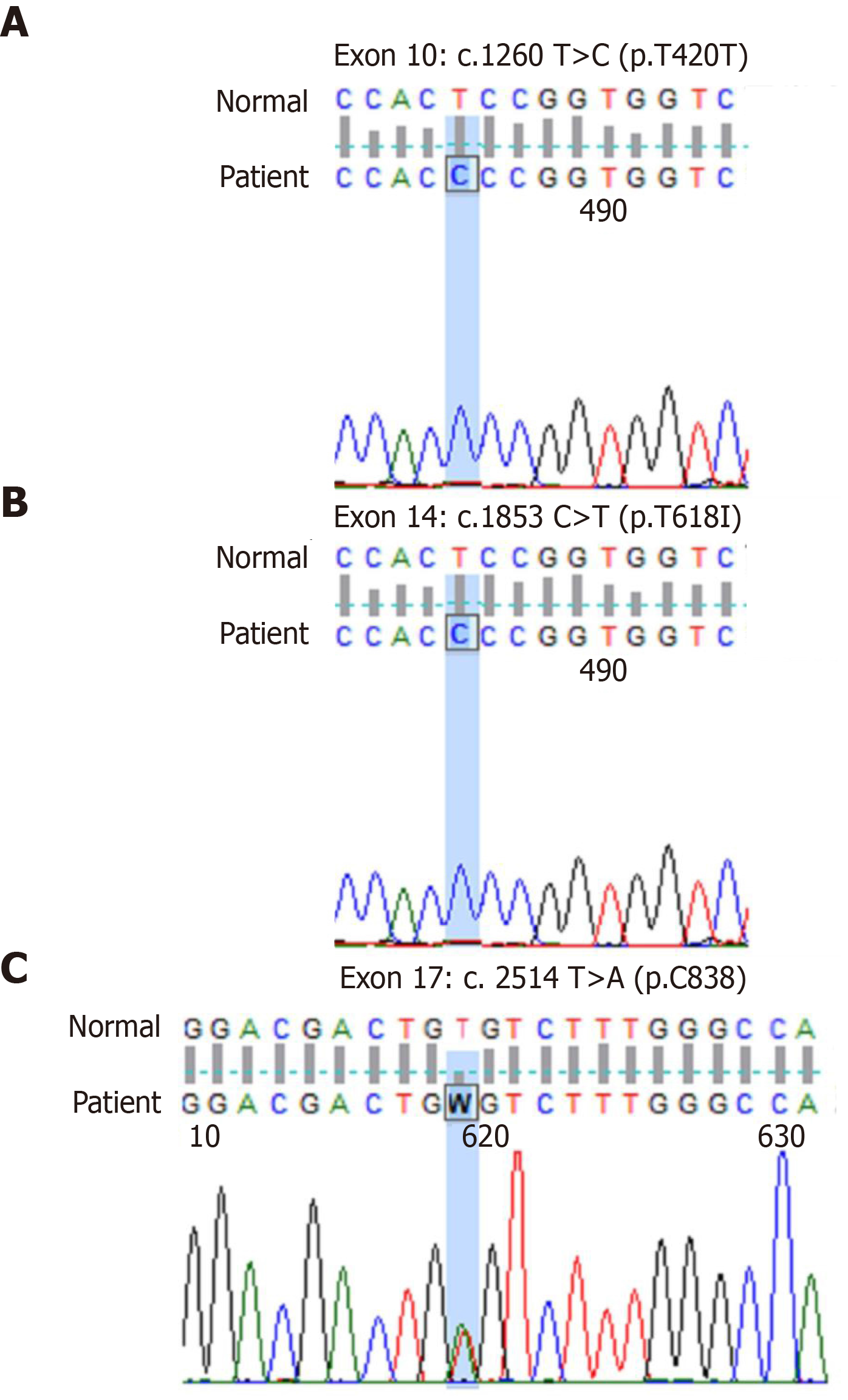
Figure 3 Sequencing of mutant sites.
A: c.1260T>C (p.T420T) mutation; B: c.1853C>T (p.T618I) mutation; C: c.2514T>A (p.C838) mutation.
- Citation: Li YP, Chen N, Ye XM, Xia YS. Eighty-year-old man with rare chronic neutrophilic leukemia caused by CSF3R T618I mutation: A case report and review of literature. World J Clin Cases 2020; 8(24): 6337-6345
- URL: https://www.wjgnet.com/2307-8960/full/v8/i24/6337.htm
- DOI: https://dx.doi.org/10.12998/wjcc.v8.i24.6337


