Copyright
©The Author(s) 2020.
World J Clin Cases. Nov 6, 2020; 8(21): 5371-5379
Published online Nov 6, 2020. doi: 10.12998/wjcc.v8.i21.5371
Published online Nov 6, 2020. doi: 10.12998/wjcc.v8.i21.5371
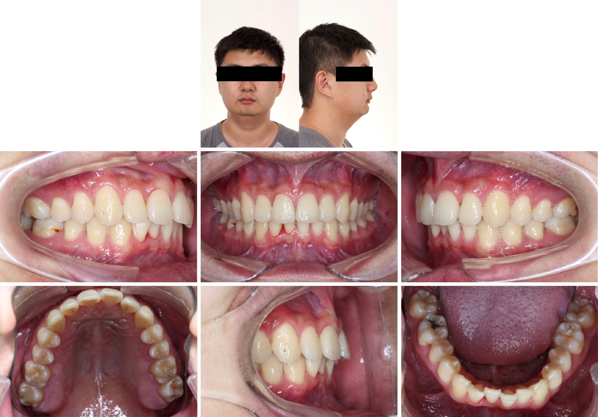
Figure 1 Pretreatment photographs.
Figure 2 Pretreatment radiographs.
A: Panoramic; B: Lateral cephalometric; C: Cephalometric measurements. ANB: Subspinale-nasion-supramental; U1: Upper first incisor; L1: Lower first incisor; MP: Mandibular plane; SN: Sella-nasion; FH: Frankfort horizontal plane; Wits: Witwatersrand.
Figure 3 Posttreatment photographs.
Figure 4 Posttreatment radiographs.
A: Panoramic; B: Lateral cephalometric; C: Cephalometric measurements. ANB: Subspinale-nasion-supramental; U1: Upper first incisor; L1: Lower first incisor; MP: Mandibular plane; SN: Sella-nasion; FH: Frankfort horizontal plane; Wits: Witwatersrand.
Figure 5 Seven-year posttreatment photographs.
- Citation: Yu TT, Li J, Liu DW. Seven-year follow-up of the nonsurgical expansion of maxillary and mandibular arches in a young adult: A case report. World J Clin Cases 2020; 8(21): 5371-5379
- URL: https://www.wjgnet.com/2307-8960/full/v8/i21/5371.htm
- DOI: https://dx.doi.org/10.12998/wjcc.v8.i21.5371


