Copyright
©The Author(s) 2019.
World J Clin Cases. Feb 6, 2019; 7(3): 291-299
Published online Feb 6, 2019. doi: 10.12998/wjcc.v7.i3.291
Published online Feb 6, 2019. doi: 10.12998/wjcc.v7.i3.291
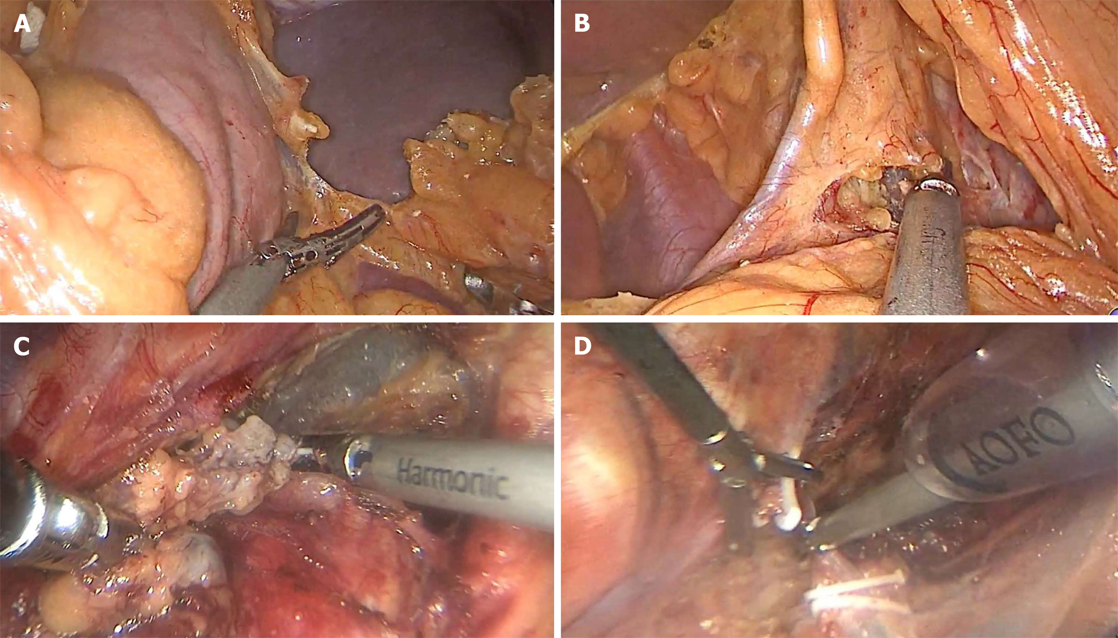
Figure 1 Surgical procedure of minimally invasive Ivor-Lewis esophagectomy.
A: The large curved side of the stomach was separated; B: The small curved omentum of the stomach was separated; C: The lymph nodes near the right recurrent laryngeal nerve were cleared; D: The odd vein bow was clipped and severed.
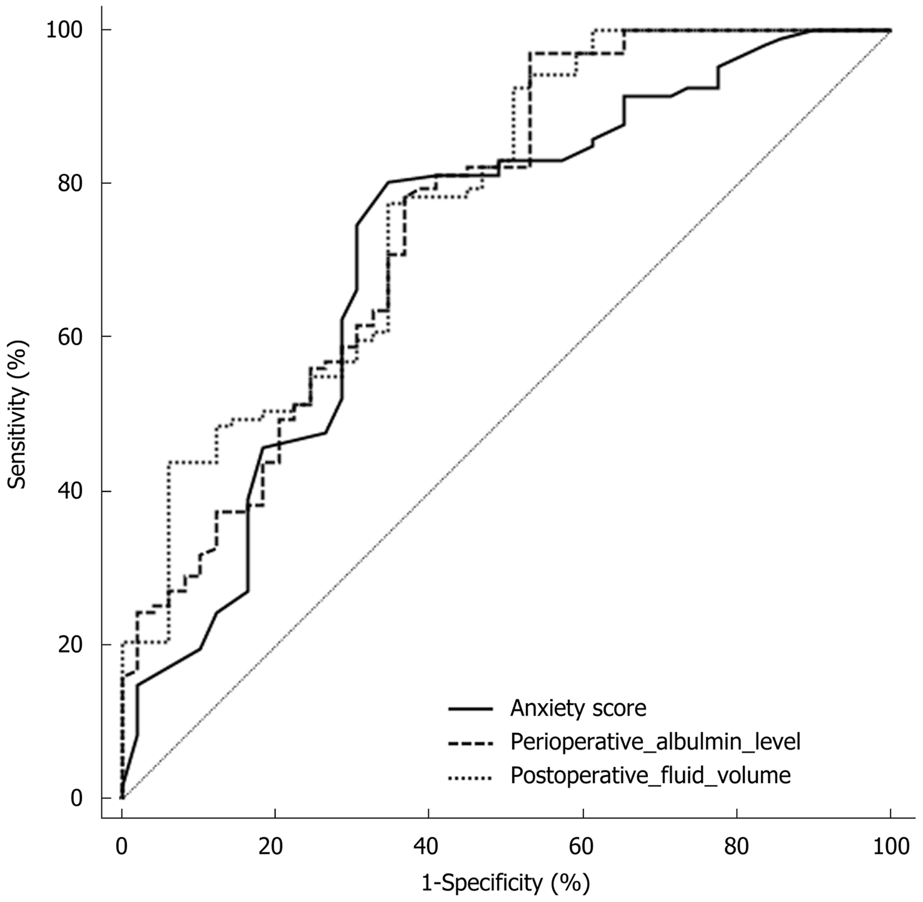
Figure 2 Receiver operating characteristic curve analysis for predicting postoperative early delayed gastric emptying in esophageal cancer patients.
- Citation: Huang L, Wu JQ, Han B, Wen Z, Chen PR, Sun XK, Guo XD, Zhao CM. Influencing factors of postoperative early delayed gastric emptying after minimally invasive Ivor-Lewis esophagectomy. World J Clin Cases 2019; 7(3): 291-299
- URL: https://www.wjgnet.com/2307-8960/full/v7/i3/291.htm
- DOI: https://dx.doi.org/10.12998/wjcc.v7.i3.291


