Copyright
©The Author(s) 2019.
World J Clin Cases. Oct 6, 2019; 7(19): 3062-3068
Published online Oct 6, 2019. doi: 10.12998/wjcc.v7.i19.3062
Published online Oct 6, 2019. doi: 10.12998/wjcc.v7.i19.3062
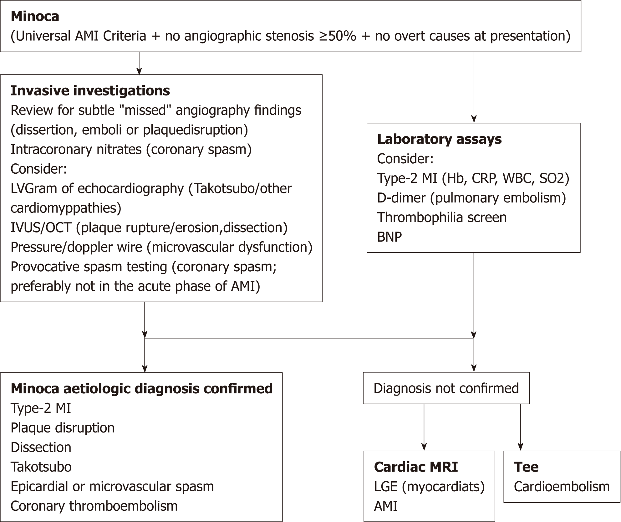
Figure 1 European Society of Cardiology working group position paper on myocardial infarction with nonobstructive coronary arteries.
MINOCA: Myocardial infarction with nonobstructive coronary arteries; BNP: Brain natriuretic peptide; CRP: C-reactive protein; WBC: White blood cells; AMI: Acute myocardial infarction.
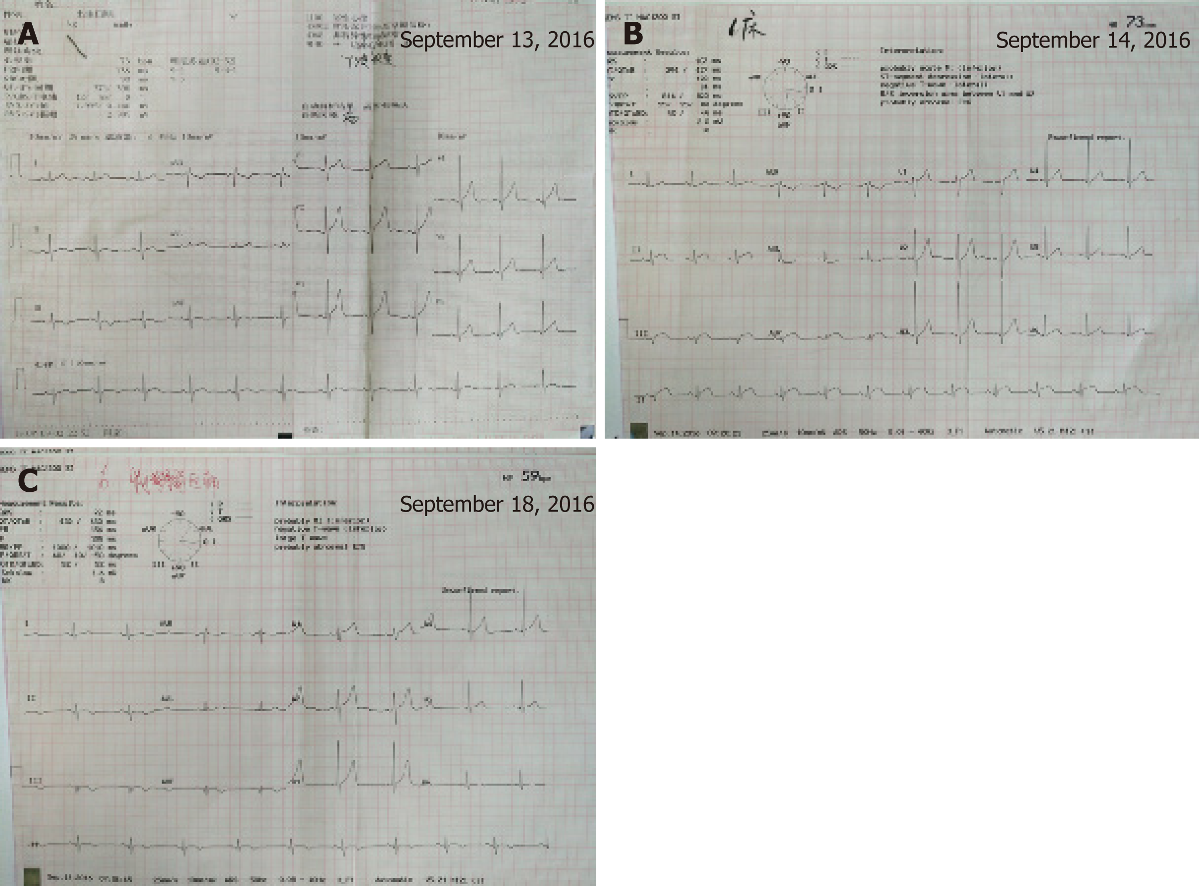
Figure 2 Changes in electrocardiography from September 13 to 18 during the acute myocardial infarction.
A: Electrocardiography on September 13, 2016; B: Electrocardiography on September 14, 2016; C: Electrocardiography on September 18, 2016.
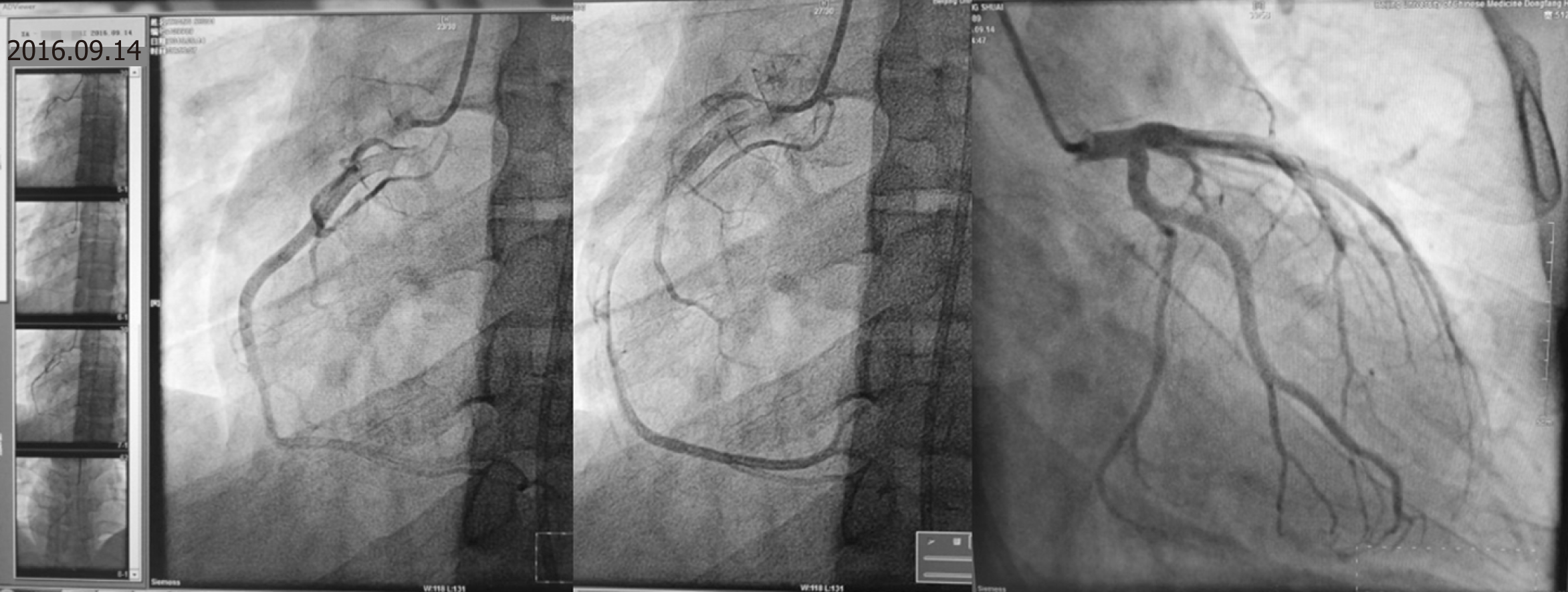
Figure 3 Results of coronary angiography.
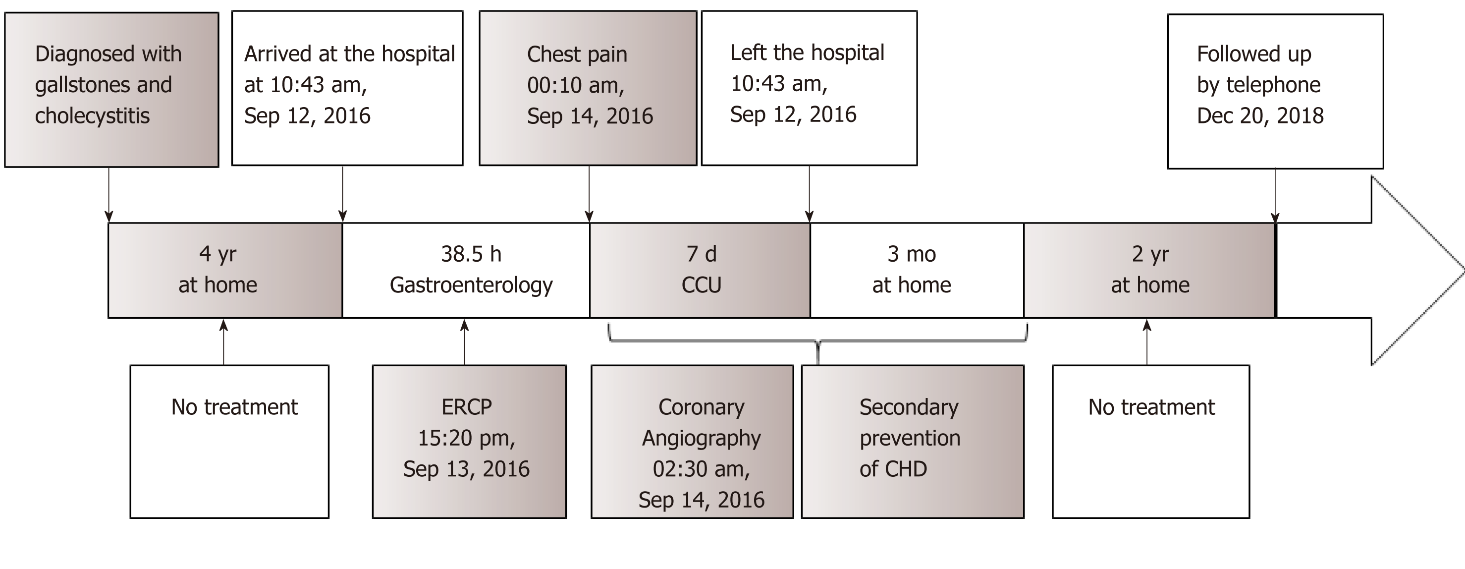
Figure 4 Timeline of the patient.
CCU: Coronary care unit; CHD: Coronary heart disease; ERCP: Endoscopic retrograde cholangiopancreatography.
- Citation: Li D, Li Y, Wang X, Wu Y, Cui XY, Hu JQ, Li B, Lin Q. Diagnosis of myocardial infarction with nonobstructive coronary arteries in a young man in the setting of acute myocardial infarction after endoscopic retrograde cholangiopancreatography: A case report. World J Clin Cases 2019; 7(19): 3062-3068
- URL: https://www.wjgnet.com/2307-8960/full/v7/i19/3062.htm
- DOI: https://dx.doi.org/10.12998/wjcc.v7.i19.3062


