Copyright
©The Author(s) 2019.
World J Clin Cases. Jul 6, 2019; 7(13): 1703-1710
Published online Jul 6, 2019. doi: 10.12998/wjcc.v7.i13.1703
Published online Jul 6, 2019. doi: 10.12998/wjcc.v7.i13.1703
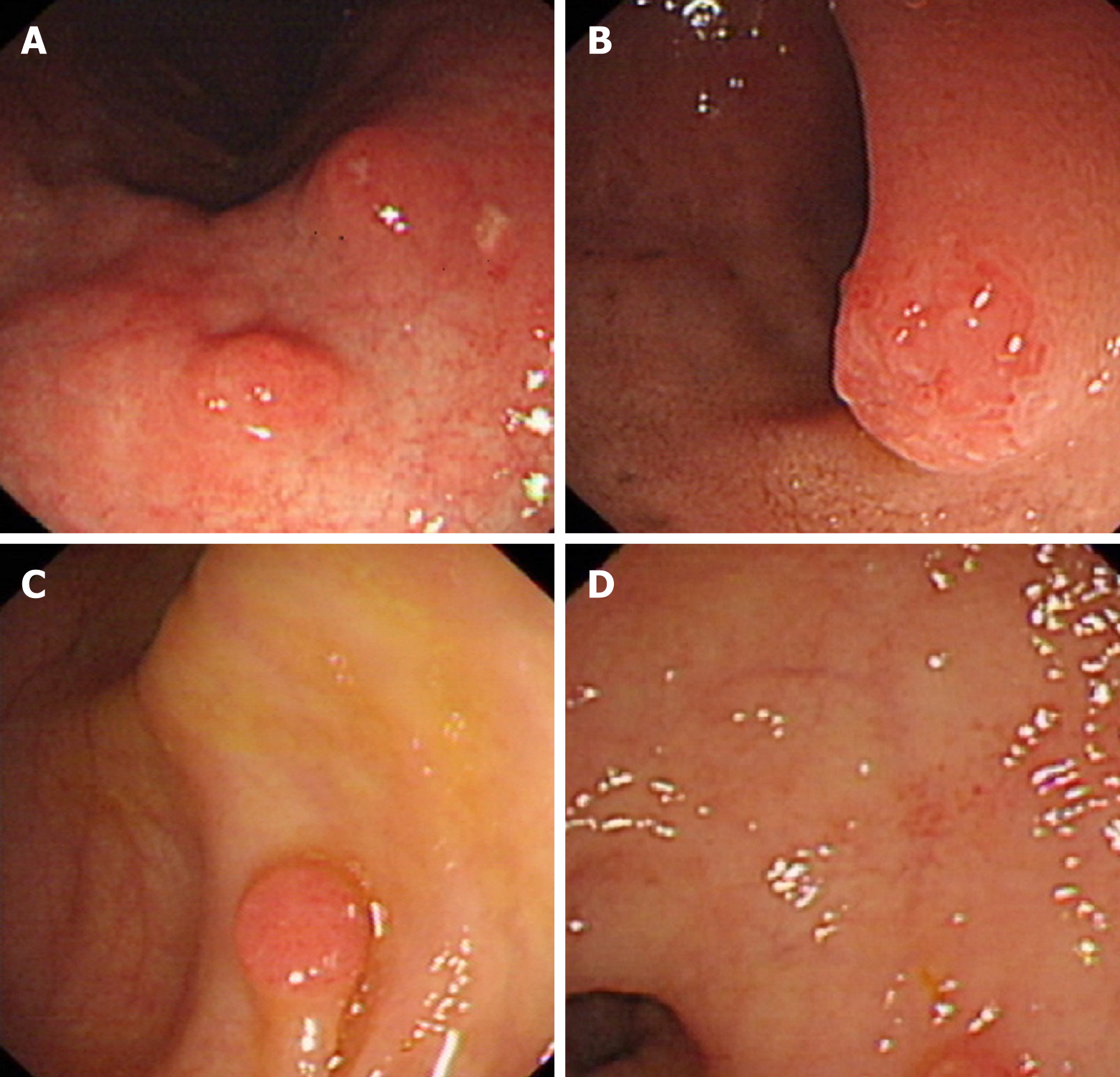
Figure 1 Endoscopy results.
Multiple small polypoid lesions without stalk were observed in A: The stomach; B: The duodenum; C: The colon; D: The rectum.
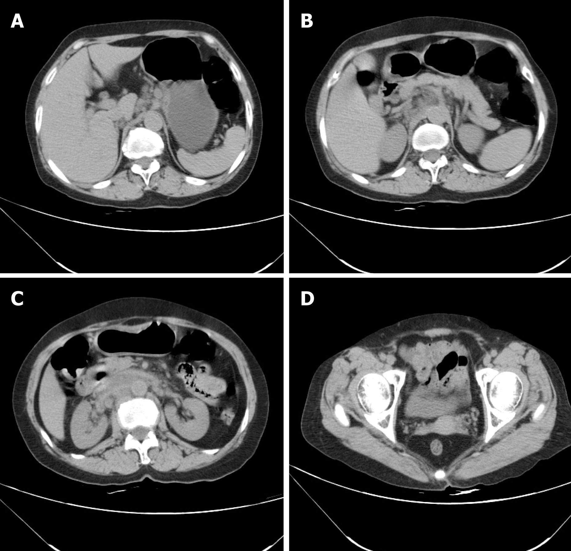
Figure 2 Computed tomography findings.
A-D: Computed tomography images not revealing substantial evidence for the existance of malignant lesions in the gastrointestinal wall.
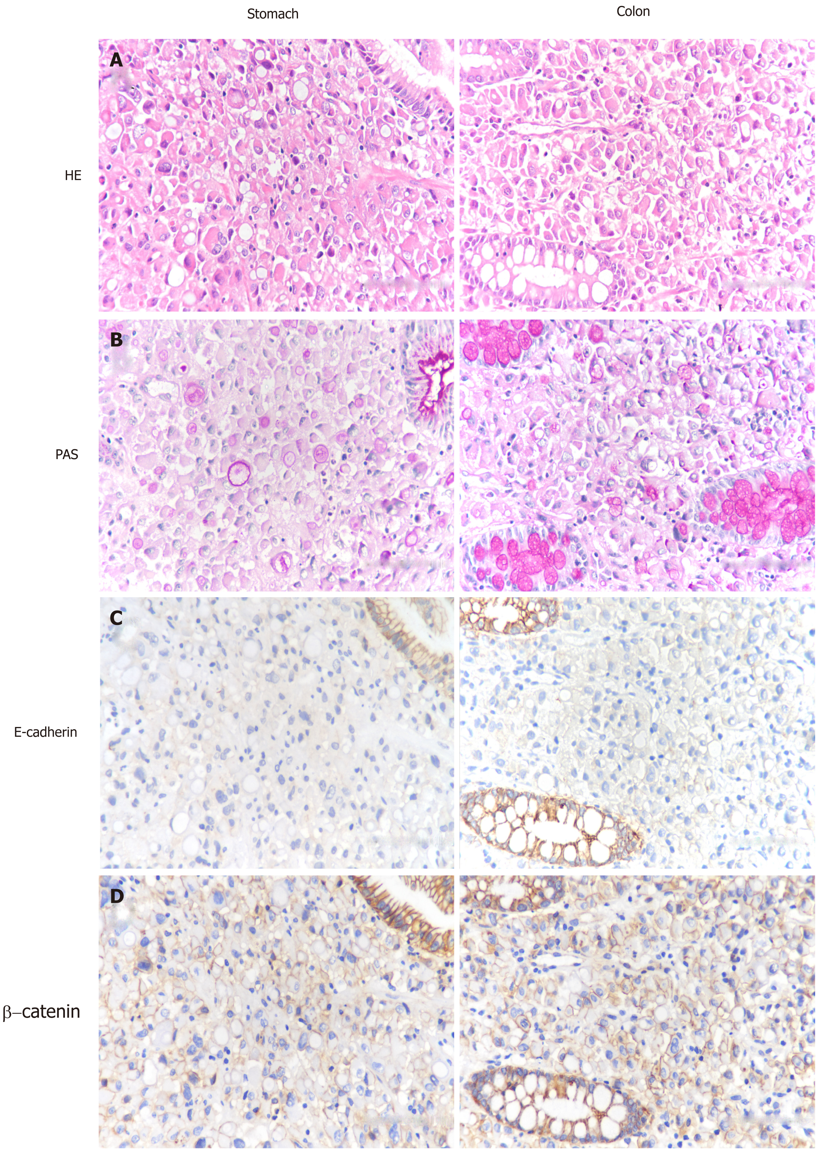
Figure 3 Morphology and immunophenotype of the lesion.
A: H&E staining showed poorly differentiated signet ring carcinoma; B: Tumor cells were negative for E-cadherin; C: β-catenin was translocated from the membrane to the cytoplasm and nucleus in malignant cells; D: Periodic acid Schiff (PAS) staining revealed the presence of mucin in the cytoplasm of signet ring cells. Original magnification, ×400.
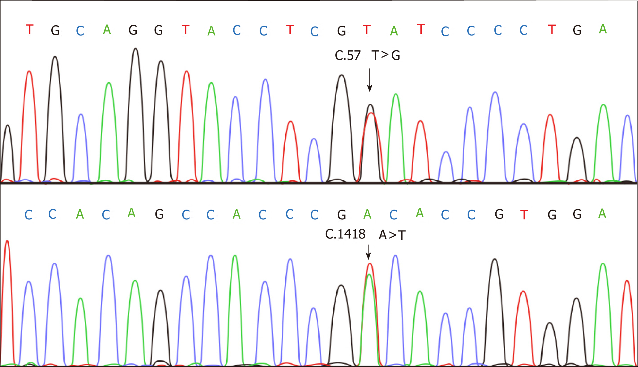
Figure 4 CDH1 sequencing.
Two base substitutions were confirmed in the CDH1 gene: C.57 T>G and C.1418 A>T.
- Citation: Hu MN, Lv W, Hu RY, Si YF, Lu XW, Deng YJ, Deng H. Synchronous multiple primary gastrointestinal cancers with CDH1 mutations: A case report. World J Clin Cases 2019; 7(13): 1703-1710
- URL: https://www.wjgnet.com/2307-8960/full/v7/i13/1703.htm
- DOI: https://dx.doi.org/10.12998/wjcc.v7.i13.1703


