Copyright
©The Author(s) 2018.
World J Clin Cases. Nov 26, 2018; 6(14): 825-829
Published online Nov 26, 2018. doi: 10.12998/wjcc.v6.i14.825
Published online Nov 26, 2018. doi: 10.12998/wjcc.v6.i14.825
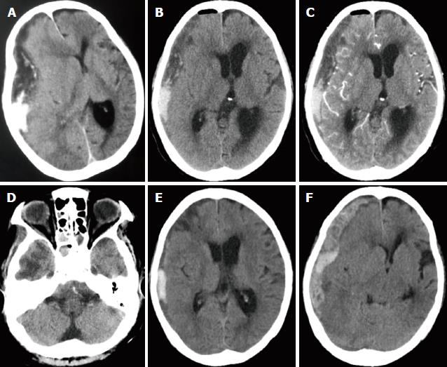
Figure 1 Radiographic images of the presenting case.
A: Preoperative unenhanced CT revealed a hypodense crescent-shaped extra-axial fluid collection mixed with a local hyperdense lesion over the right hemisphere and marked midline shift of the brain; B: Immediate postoperative unenhanced CT scan demonstrated a slightly decreased subdural empyema and retraction of middle structures of the brain; C: An enhanced CT scan which is corresponding to figure B showed no enhancement of the thick rim or adjacent cerebral cortex; D: Hypointense areas (finger-like) were seen in the right lower temporal lobe; E: Unenhanced CT scan indicating significantly decreased subdural empyema at day 4; F: Unenhanced CT scan showed newly subdural hemorrhage and the middle shift of brain structures at day 5.
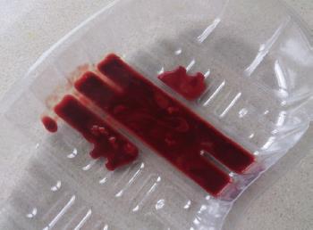
Figure 2 Drainage showed pus mixed with blood.
- Citation: Chen JC, Tan DH, Xue ZB, Yang SY, Li Y, Lai RL. Subdural empyema complicated with intracranial hemorrhage in a postradiotherapy nasopharyngeal carcinoma patient: A case report and review of literature. World J Clin Cases 2018; 6(14): 825-829
- URL: https://www.wjgnet.com/2307-8960/full/v6/i14/825.htm
- DOI: https://dx.doi.org/10.12998/wjcc.v6.i14.825


