Copyright
©The Author(s) 2018.
World J Clin Cases. Nov 26, 2018; 6(14): 811-819
Published online Nov 26, 2018. doi: 10.12998/wjcc.v6.i14.811
Published online Nov 26, 2018. doi: 10.12998/wjcc.v6.i14.811
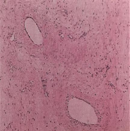
Figure 1 Pathological results after H-E staining of one case.
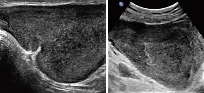
Figure 2 Gray-scale ultrasonic images showing irregular hypoechoic masses with internal echogenicity and well-defined margins.
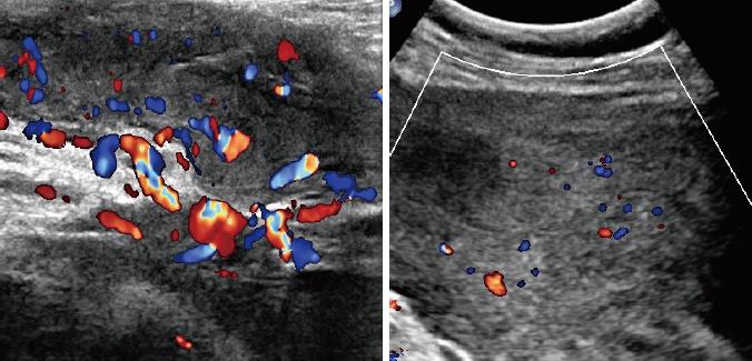
Figure 3 Internal blood flows in the mass detected by colour Doppler ultrasound.
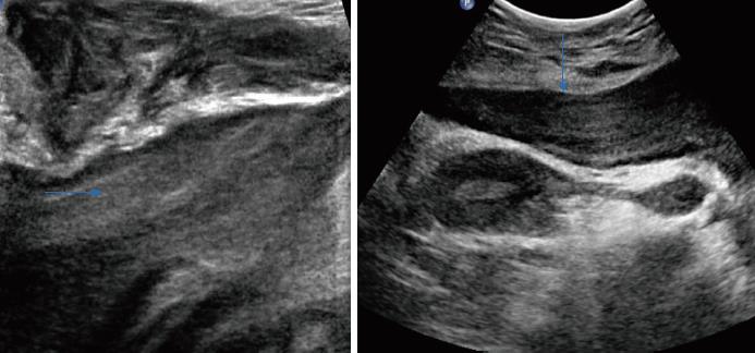
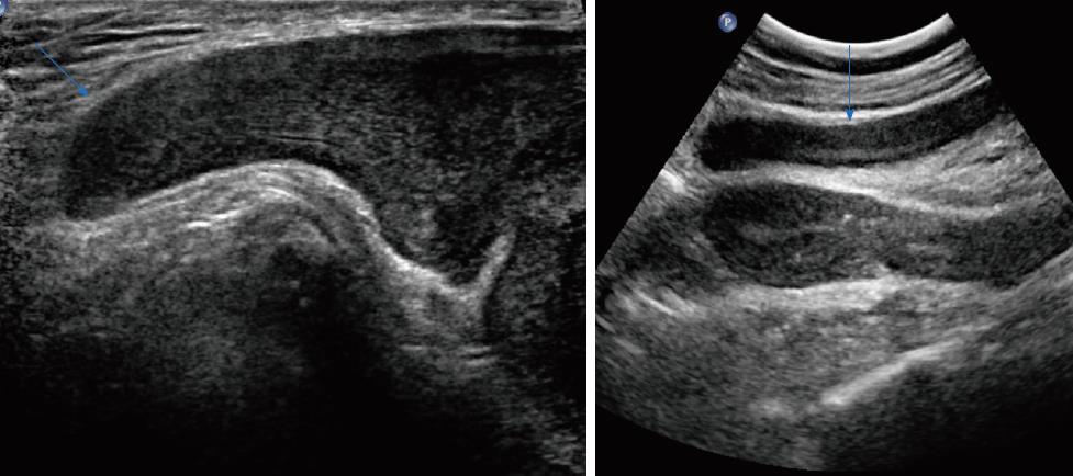
Figure 4 Laminated or swirled appearance of inner echogenicity (blue arrows).
Figure 5 The finger-like growth pattern in the border of the mass could be demonstrated in both cases (blue arrows).
Figure 6 Magnetic resonance imaging images of case 4.
A: Intermediate-intensity signals on TI-weighted sequence; B: Intermediate to high-intensity signals on T2-weighted sequence; C: Rapid and uneven enhancement pattern was shown after the injection of contrast agent.
- Citation: Zhao CY, Su N, Jiang YX, Yang M. Application of ultrasound in aggressive angiomyxoma: Eight case reports and review of literature. World J Clin Cases 2018; 6(14): 811-819
- URL: https://www.wjgnet.com/2307-8960/full/v6/i14/811.htm
- DOI: https://dx.doi.org/10.12998/wjcc.v6.i14.811


