Copyright
©The Author(s) 2017.
World J Clin Cases. Jan 16, 2017; 5(1): 9-13
Published online Jan 16, 2017. doi: 10.12998/wjcc.v5.i1.9
Published online Jan 16, 2017. doi: 10.12998/wjcc.v5.i1.9
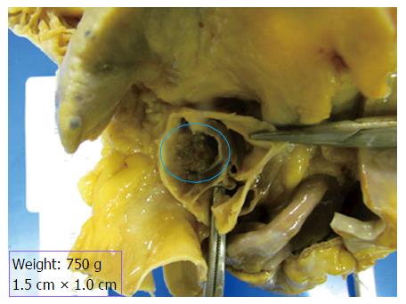
Figure 1 Autopsy specimen of the heart demonstrating a cardiac papillary fibroelastoma on the aortic valve.
Note the large bulky tan-white non-encapsulated, rubbery, firm lesion with short pedicle and multiple papillary fronds on the aortic valve measuring 1.5 cm × 1.0 cm in size, completely occluding the right coronary ostium (blue circle).
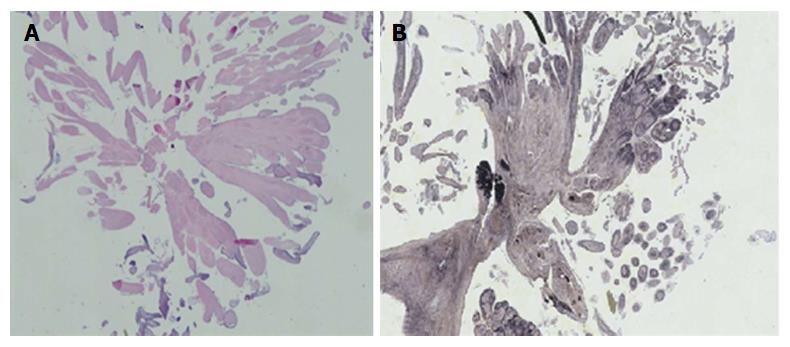
Figure 2 Microscopic photograph of the cardiac papillary fibroelastoma.
A: Histological section of the mass shows benign papillary lesion comprised of a single layer of endocardial cells overlies a thin layer of mucopolysaccharide matrix and underlying, almost acellular, avascular stroma composed predominantly of elastic fibers; B: Elastin stain reveals concentric elastin fibres within the papillary excrescences.
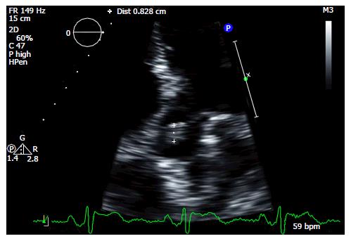
Figure 3 Transthoracic echocardiogram showing a cardiac papillary fibroelastoma on the aortic valve (marked).
- Citation: Yandrapalli S, Mehta B, Mondal P, Gupta T, Khattar P, Fallon J, Goldberg R, Sule S, Aronow WS. Cardiac papillary fibroelastoma: The need for a timely diagnosis. World J Clin Cases 2017; 5(1): 9-13
- URL: https://www.wjgnet.com/2307-8960/full/v5/i1/9.htm
- DOI: https://dx.doi.org/10.12998/wjcc.v5.i1.9


