Copyright
©The Author(s) 2017.
World J Clin Cases. Jan 16, 2017; 5(1): 18-23
Published online Jan 16, 2017. doi: 10.12998/wjcc.v5.i1.18
Published online Jan 16, 2017. doi: 10.12998/wjcc.v5.i1.18
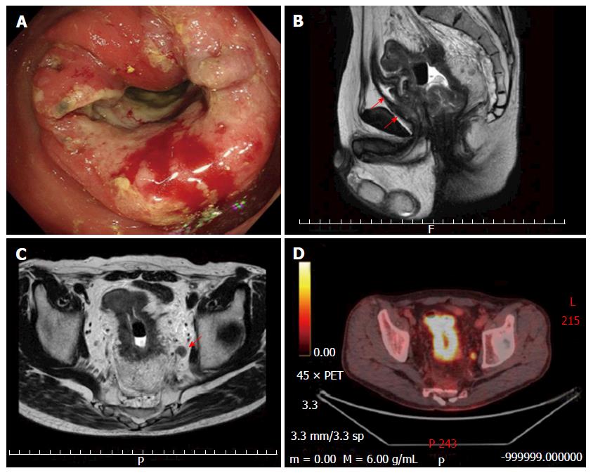
Figure 1 Evaluation of clinical findings.
Colonoscopy showed a circumferential mass at the lower rectum (A); Sagittal magnetic resonance imaging (MRI) of the pelvis showed rectal mass with involvement prostate and seminal vesicles (red arrows) (B), and perirectal fat (C); The enlarged lymph node in the left obturator detected by coronal MRI (red arrow) showed obvious metabolically active foci 18-fluorodeoxyglucose-positron emission tomography/computed tomography evaluation (D).
Figure 2 Rectal tumor perforation suggestive of chemoradiationdamage.
Radiotherapy was delivered to the whole pelvis through three (one posterior-anterior and two lateral) or four (one anterior-posterior, one posterior-anterior and two lateral) fields using a 10-MV linear accelerator in the prone position (A); Coronal computed tomography findings showed a small bubble of extra-luminal gas (red arrow) (B); Preoperative colonoscopicfindings for radical surgery showed excavation with mucosa necrosis (red arrow) suggestive of chemoradiationdamage in the rectal tumor (C).
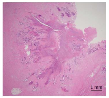
Figure 3 Histological findings of the resected specimen showed a wide field of tumor necrosis with fibril formation (H-E stain).
- Citation: Takase N, Yamashita K, Sumi Y, Hasegawa H, Yamamoto M, Kanaji S, Matsuda Y, Matsuda T, Oshikiri T, Nakamura T, Suzuki S, Koma YI, Komatsu M, Sasaki R, Kakeji Y. Local advanced rectal cancer perforation in the midst of preoperative chemoradiotherapy: A case report and literature review. World J Clin Cases 2017; 5(1): 18-23
- URL: https://www.wjgnet.com/2307-8960/full/v5/i1/18.htm
- DOI: https://dx.doi.org/10.12998/wjcc.v5.i1.18


