Copyright
©The Author(s) 2017.
World J Clin Cases. Jan 16, 2017; 5(1): 1-8
Published online Jan 16, 2017. doi: 10.12998/wjcc.v5.i1.1
Published online Jan 16, 2017. doi: 10.12998/wjcc.v5.i1.1
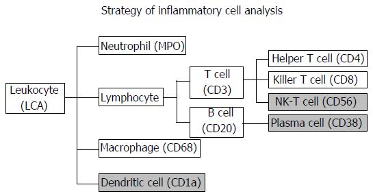
Figure 1 The inflammatory cell analysis strategy.
Gray filled shows items which was not able to analyze by Tissue studio (because of positive stain of tumor cells). LCA: Leukocyte common antigen; MPO: Myeloperoxidase.
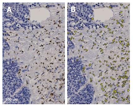
Figure 2 Images of CD8+ lymphocytes.
A: Original image of CD8-immunostained slide of an invasive area; B: The software Tissue Studio detects the positive cells and counts them automatically.
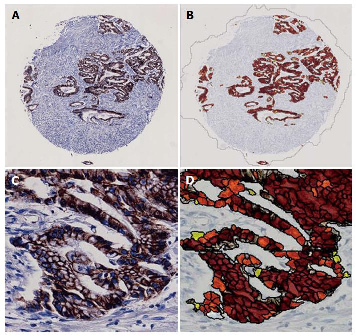
Figure 3 Images of E-cadherin expression in cancer cells.
A: Low-magnification image of an E-cadherin-immunostained TMA core; B: Low-magnification image of E-cadherin-immunostained TMA core analyzed by Tissue Studio; C: High-magnification (× 400) image of an E-cadherin-immunostained TMA core; D: High-magnification (× 400) image of an E-cadherin-immunostained TMA core analyzed by Tissue Studio. The membranous expression of E-cadherin was assigned to three categories of the degree of expression: High (brown), medium (orange) and low (yellow). The histological score was calculated by the following formula: 1 × %Low + 2 × %Medium + 3 × %High. TMA: Tissue microarray.
Figure 4 The single linear regression models of the relationship between E-cadherin expression (histological score) and the numbers of cells.
The cells were positively immunostained by LCA (A), CD3 (B) and CD8 (C) antibodies.
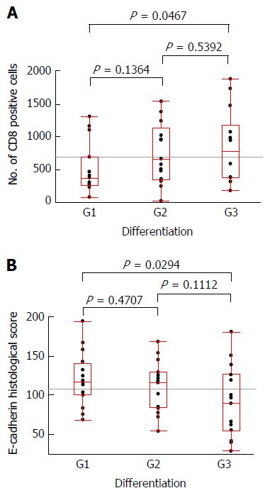
Figure 5 The box-and-whisker plots regarding pathologically assessed differentiation and the number of CD8+ cells (A) and the E-cadherin histological score (B).
Significant differences were observed in the comparison of G1 and G3 cases according to the number of CD8+ cells (P = 0.0467) and according to the E-cadherin histological score (P = 0.0294), respectively.
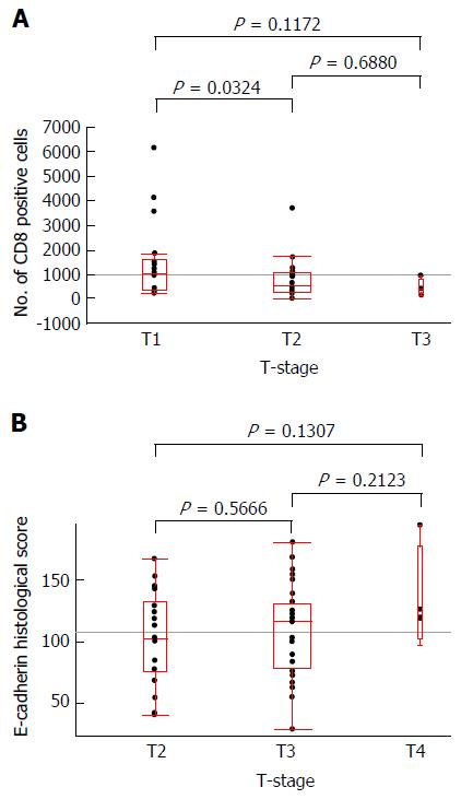
Figure 6 The box-and-whisker plots regarding T-stage and the number of CD8+ cells (A) and the E-cadherin histological score (B).
A significant difference was observed in the comparison of T2 and T3 cases according to the number of CD8+ cells (P = 0.0324).
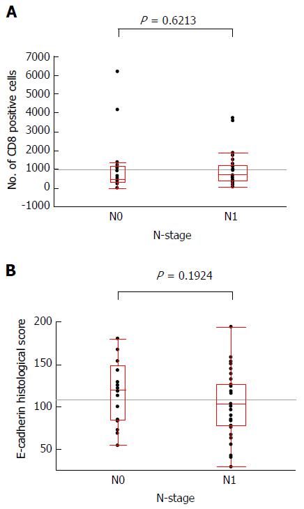
Figure 7 The box-and-whisker plots regarding N-stage and the number of CD8+ cells (A) and the E-cadherin histological score (B).
No significant difference was observed in each comparison.
- Citation: Kai K, Masuda M, Aishima S. Inverse correlation between CD8+ inflammatory cells and E-cadherin expression in gallbladder cancer: Tissue microarray and imaging analysis. World J Clin Cases 2017; 5(1): 1-8
- URL: https://www.wjgnet.com/2307-8960/full/v5/i1/1.htm
- DOI: https://dx.doi.org/10.12998/wjcc.v5.i1.1


