Copyright
©The Author(s) 2016.
World J Clin Cases. Jan 16, 2016; 4(1): 25-29
Published online Jan 16, 2016. doi: 10.12998/wjcc.v4.i1.25
Published online Jan 16, 2016. doi: 10.12998/wjcc.v4.i1.25
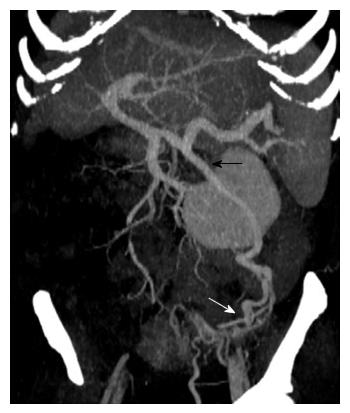
Figure 1 Computed tomography-scan three-dimensional reconstruction in maximum intensity projection showed the ectopic varices (white arrow) at the colostomy conduit fed by the inferior mesenteric vein (black arrow).
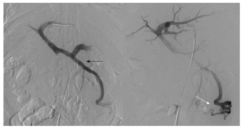
Figure 2 Digital subtraction venography showed hepatofugal flow toward the stoma by inferior mesenteric vein (black arrow) feeding the stomal varices (white arrow).
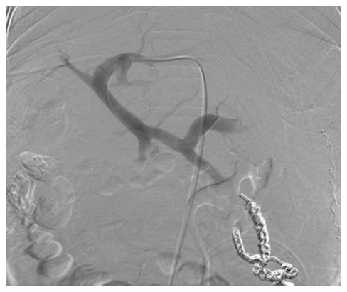
Figure 3 Digital subtraction venography after embolization demonstrated complete obliteration of variceal branches.
- Citation: Maciel MJS, Pereira OI, Motta Leal Filho JM, Ziemiecki Junior E, Cosme SL, Souza MA, Carnevale FC. Peristomal variceal bleeding treated by coil embolization using a percutaneous transhepatic approach. World J Clin Cases 2016; 4(1): 25-29
- URL: https://www.wjgnet.com/2307-8960/full/v4/i1/25.htm
- DOI: https://dx.doi.org/10.12998/wjcc.v4.i1.25


