Copyright
©The Author(s) 2015.
World J Clin Cases. May 16, 2015; 3(5): 470-473
Published online May 16, 2015. doi: 10.12998/wjcc.v3.i5.470
Published online May 16, 2015. doi: 10.12998/wjcc.v3.i5.470
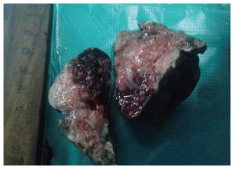
Figure 1 Congenital pulmonary airway malformation Type III-Cut section of lung showing solid areas with few slit like spaces with focal areas of haemorrhages.
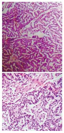
Figure 2 Photomicrograph showing congenital pulmonary airway malformation Type III with pneumonia.
Neutrophilic infiltrate in alveoli and cystic spaces. (HE × high power).
Figure 3 Photomicrograph showing congenital pulmonary airway malformation Type II-Bronchiole like structures are lined by cuboidal to columnar epithelium with back-to-back arrangement (HE × high power).
- Citation: Bolde S, Pudale S, Pandit G, Ruikar K, Ingle SB. Congenital pulmonary airway malformation: A report of two cases. World J Clin Cases 2015; 3(5): 470-473
- URL: https://www.wjgnet.com/2307-8960/full/v3/i5/470.htm
- DOI: https://dx.doi.org/10.12998/wjcc.v3.i5.470


